
Taking the advantage of high spatial coherence and brightness given by the liquid metal jet anode tube, the scientists at university of Göttingen have succeeded in demonstrating the X-ray phase-contrast tomography as a novel, non-invasive imaging methodology to do 3-D virtual histology at isotropic sub-cellular resolution. With the optimized system, image reconstruction and analysis procedures, the 3-D cytoarchitecture of the unstained paraffin-embedded human cerebellum has been studied and compared with setups at both synchrotron radiation and laboratory source. The latter, which has revealed the location of neurons and is in good analog to those from synchrotron, will endorse broader applications with good accessibility, sufficient spatio-temporal resolution and large field-of-view.
Related Posts
Decision-quality fast CT for battery inspection
Steffen Masuch and Klaus Dröder, TU Braunschweig; and Sophie Gräfnitz, PowerCo.
Ingrid AksnesMarch 26, 2026

Extremely high-speed X-ray radiography at micrometer resolution to reveal hidden dynamics for failure and root cause analysis of electronics
Julius Hållstedt, Emil Espes, Till Dreier, and Daniel Nilsson, Excillum; Spyridon Gkoumas, DECTRIS Ltd.
Ingrid AksnesDecember 2, 2025
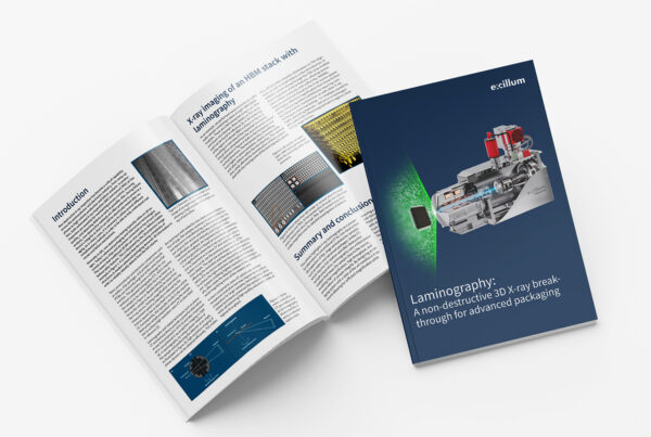
Laminography: A non-destructive 3D X-ray breakthrough for advanced packaging
Till Dreier and Julius Hållstedt, Excillum.
Ingrid AksnesOctober 8, 2025