Kristina Kutukova, Bartlomiej Lechowski, Joerg Grenzer, Peter Krueger, André Clausner, and Ehrenfried Zschech
Nanomaterials 2024, 14, 448.
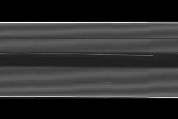
High-resolution imaging of Cu/low-k on-chip interconnect stacks in advanced microelectronic products is demonstrated using full-field transmission X-ray microscopy (TXM). The comparison of two lens-based laboratory X-ray microscopes that are operated at two different photon energies, 8.0 keV and 9.2 keV, shows a contrast enhancement for imaging of copper nanostructures embedded in insulating organosilicate glass of a factor of 5 if 9.2 keV photons are used. Photons with this energy (Ga-Kα radiation) are generated from a Ga-containing target of a laboratory X-ray source applying the liquid-metal-jet technology. The 5 times higher contrast compared to the use of Cu-Kα radiation (8.0 keV photon energy) from a rotating anode X-ray source is caused by the fact that the energy of the Ga-Kα emission line is slightly higher than that of the Cu-K absorption edge (9.0 keV photon energy). The use of Ga-Kα radiation is of particular advantage for imaging of copper interconnects with dimensions from several 100 nm down to several 10 nm in a Cu/SiO2 or Cu/low-k backend-of-line stack. Physical failure analysis and reliability engineering in the semiconductor industry will benefit from high-contrast X-ray images of sub-μm copper structures in microchips.