Nano-CT/micro-CT
The technology development towards fast high-resolution imaging has been driven by the needs in scientific research, industrial R&D, and production quality control. To visualize fine details of the microstructure in the object, the imaging can be done either by using X-ray radiation coming from a small emission spot, or by using X-ray optics to build a microscope setup.

An X-ray tube with extremely small emission spot size can be used for high resolution imaging without optics. The advantages of this approach without optics are the efficiency across the full energy spectrum as well as the ease in getting a large field of view. Thanks to the geometric magnification produced by the point source, the object can be imaged at much higher resolution than the pixel size of the detector.
With an industry-leading emission spot size down to 300 nm, the Excillum NanoTube N3 enables lensless sub-micron X-ray microscopy and Nano-CT in the laboratory with down to 150 nm resolution. For the applications where a 5-20 µm spot is sufficient, the MetalJet offers up to 17 times more brightness than any other microfocus tube.
High resolution imaging can sometimes require very long acquisition times. During the acquisition, high stability is necessary in all parts of the imaging system. The source emission spot stability is critical, in terms of spot size change and positional drift, since it may cause image blurring or require additional efforts in image processing.
The high flux and exceptional spot stability of the NanoTube N3 makes it ideal for demanding 3D computed tomography applications as well as 2D imaging in both industrial and research environments.
Application examples for Nano-CT/micro-CT
CT for NDT, metrology and inspection
In-line micro-CT
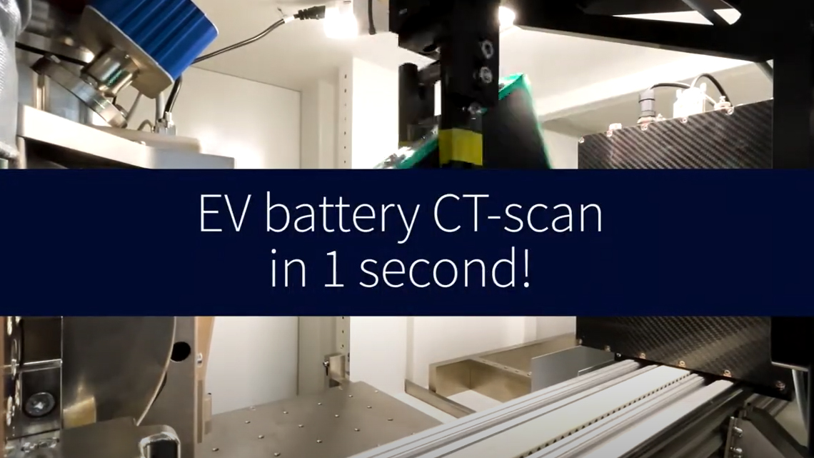
With the MetalJet E1+, Excillum is looking to enable high-speed 3D X-ray inspection of industrial samples, such as batteries. In this video, you’ll see an example of how we achieve a full CT-scan with micrometer resolution of an electrolyte filled battery cell, taken from a VW Golf GTE, in only 1 second.
100 % 3D X-ray inspection, or 3D complement to 2D inspection in unclear cases, is a promising path to satisfactory quality control. But, in order to achieve 100 % 3D X-ray inspection, a high-power X-ray source with a micrometer-sized X-ray spot is needed – something previously not available on the market.
These experiments were performed at Excillum’s facilities in Sweden using our MetalJet E1+, a high-performance detector from Direct Conversion (Thor FX20.256 CdTe) and a high-speed, high-precision rotation stage.
Nano-CT for electronics inspection
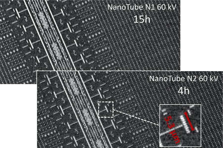
With its small spot size, the NanoTube is ideal to study the ever-shrinking structures of electronics. This example, done with predecessors of our current NanoTube N3 series, shows a tomographic slice from a nano-CT of an SD-card, performed with the NanoTube N1 60 kV (top image) and the NanoTube N2 60 kV (bottom image) – achieving a voxel sampling of 200nm.
The comparative nano-CT measurement was done by keeping a similar level of photon counts. The NanoTube N2 60 kV achieves an increase in flux by more than 3 times and, thus, the measurement time was reduced from 15 hours to 4 hours. At the same time, the signal-to-noise ratio gave even higher image quality, due to the reduced motion within the CT measurement.
A 3D rendering of the above mentioned SD card, showing the full reconstruction of the internal features and structures. Voxel sampling 200 nm.
Photo credit if not stated otherwise: Dr. Christian Fella, Fraunhofer Institute for Integrated Circuits IIS, Würzburg, Germany
Nano-CT for battery research
Nano-CT of battery cathode material
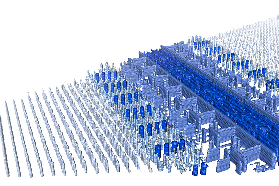
The video below shows a nano-CT of a lithium ion battery cathode material, where the microsctructure of the material can be clearly resolved. Both the aluminum cathode collector and the active LiCoO2 layer are visualized and can be analyzed further thanks to the high resolution enabled by the small X-ray spot of the Excillum NanoTube N3.
These results were generated at the applications lab at Excillum in Sweden, using our NanoTube N3 and a detector from Dectris. Visualization and analysis was made using ORS Dragonfly.
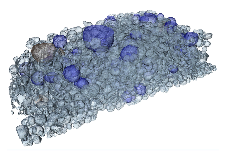
In this image, a 3D rendering of the NanoCT of a lithium ion battery cathode (NCA/LCO-E) shows particles of different sizes. Voxel sampling 140 nm.
This NanoCT system with the Excillum NanoTube has been designed, developed and commissioned at Fraunhofer IIS, Würzburg, Germany. Together with an EIGER2 CdTe detector, the system has been optimized for materials characterization and NDT applications.
Nano-CT for material sciences
A nano-CT device comprising of an Excillum NanoTube and a photon counting detector has been designed, developed and commissioned at Fraunhofer IIS, Würzburg, Germany. Together with an EIGER2 CdTe detector, the system has been optimized for materials characterization and NDT applications.
Geological and raw materials
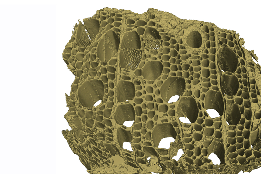
3D rendering of the phase-contrast nano-CT of an alder wood sample, revealing the inner microstructure of the wood in high resolution. Voxel sampling 300 nm.
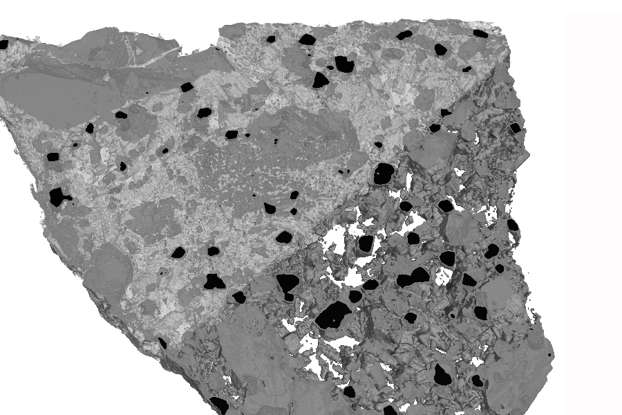
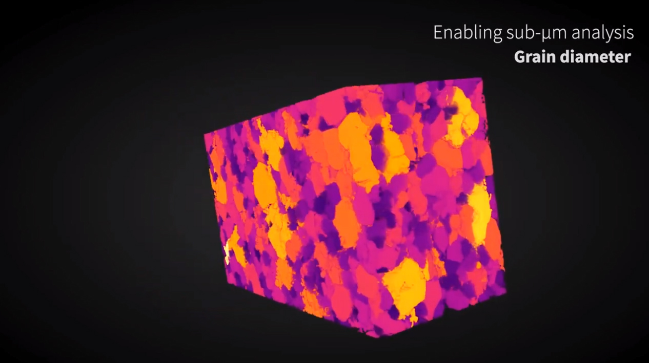
3D rendering of the phase-contrast nano-CT of a basalt sample, revealing the different phases, elements and structures help geologists to further understand this sample. Voxel sampling 350 nm.
Nano-CT for life sciences
A nano-CT device comprising of an Excillum NanoTube and a photon counting detector provides the ability of tomography with very high resolution. At the Technical University of Munich, Germany, the device has achieved a state-of-art ~100 nm spatial resolution and capability of investigating phase-contrast imaging.
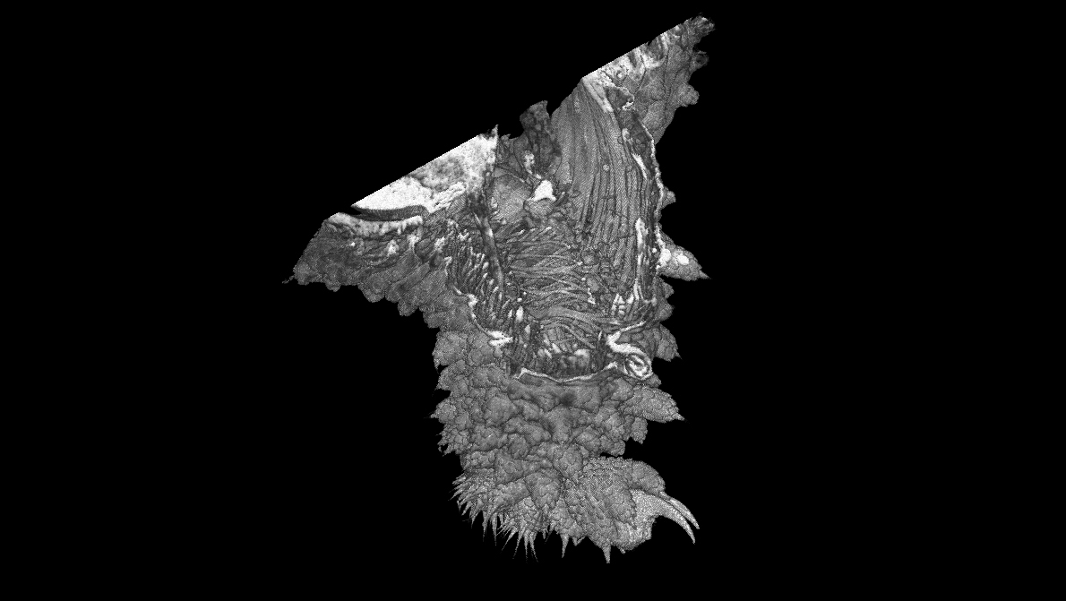
Nano-CT video and image of the limb of a velvet worm (Onychophora), 0.4 mm long. The surface morphology can be visualized with an image quality similar to scanning electron microscopy, and simultaneously the visualization of internal musculature at a resolution higher than confocal laser scanning microscopy