
An example from the Nano-CT system based on Excillum’s NanoTube at the Technical University of Munich, where the anatomical structures in cortex of the mouse kidney sample was studied with the eosin-based staining method. The compatibility of this new staining method to high resolution Micro-CT and Nano-CT have been studied. The Nano-CT images, together with the eosin-based staining method, gain a good comparison to conventional histological data, which reveal very detailed views into the 3D anatomical structure of the stained soft tissue with significantly high contrast and spatial resolution down to 200 nm. Moreover, the Nano-CT images also provide a good conjunction to the Micro-CT images to form a multi-scale CT data zooming from whole organ to small tissues. The achievement in this work indicates the future applicability of non-destructive, time-efficient virtual histology method in diagnostic screening of 3D tissue samples based on the laboratory Micro/ Nano-CT and eosin-based staining technique.
Related Posts
Decision-quality fast CT for battery inspection
Steffen Masuch and Klaus Dröder, TU Braunschweig; and Sophie Gräfnitz, PowerCo.
Ingrid AksnesMarch 26, 2026

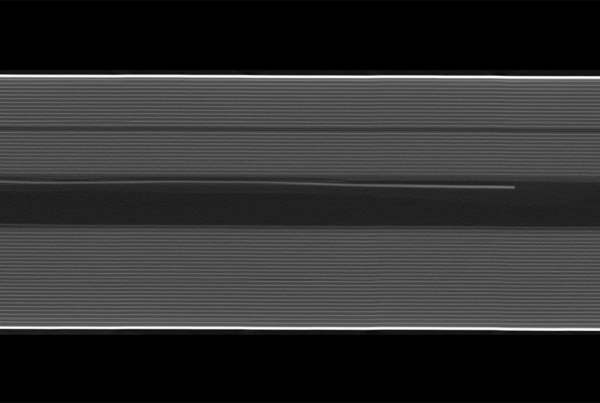
Extremely high-speed X-ray radiography at micrometer resolution to reveal hidden dynamics for failure and root cause analysis of electronics
Julius Hållstedt, Emil Espes, Till Dreier, and Daniel Nilsson, Excillum; Spyridon Gkoumas, DECTRIS Ltd.
Ingrid AksnesDecember 2, 2025
Laminography: A non-destructive 3D X-ray breakthrough for advanced packaging
Till Dreier and Julius Hållstedt, Excillum.
Ingrid AksnesOctober 8, 2025