We hope that you kicked off the new year in a brilliant way, feeling happy and energized to take on the new challenges of this year! Please, do let us know should there be anything that Excillum can do to support or assist in any of your endeavours!
We just want to take a moment to share a few highlights in and around Excillum.
Enjoy reading!
Sincerely,
Björn Hansson, CEO

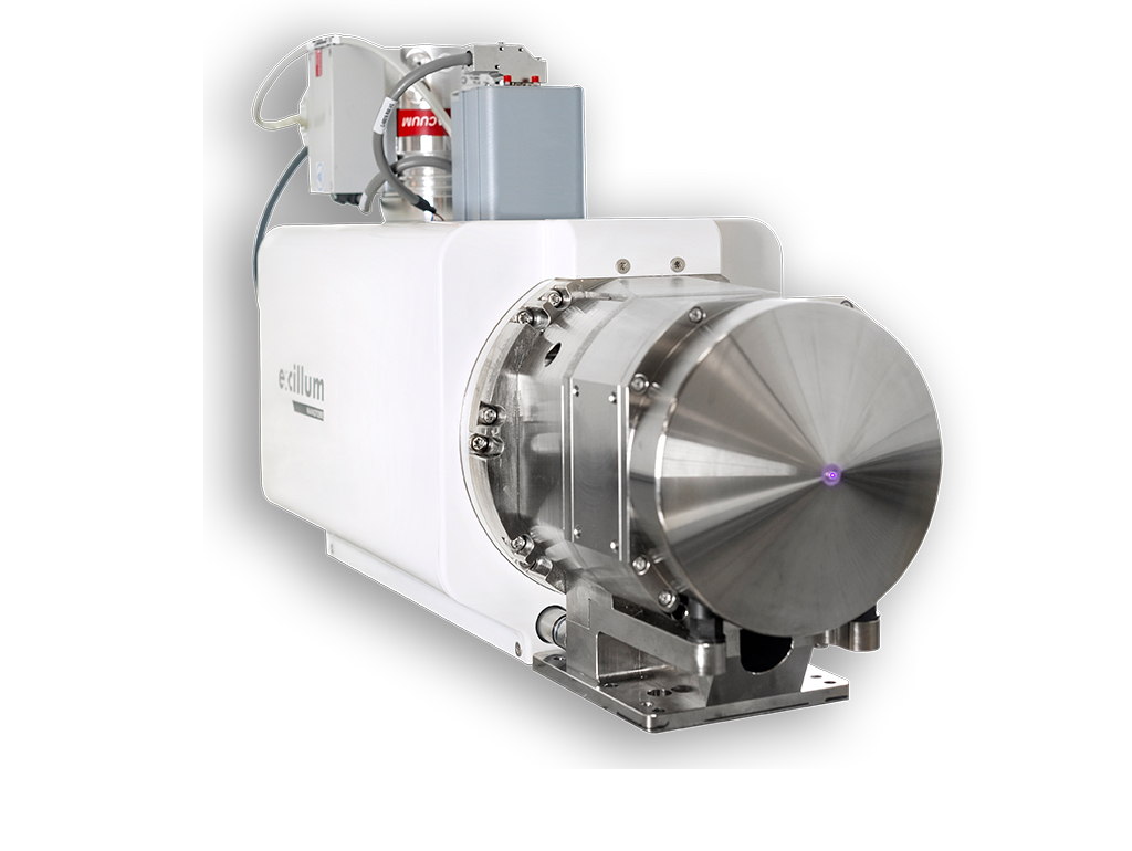
The NanoTube N1 60 kV is based on advanced electron optics and the latest tungsten-diamond transmission target technology. Automatic e-beam focusing and astigmatism correction ensures that the smallest possible, truly round spot is achieved at any voltage and current setting. The NanoTube also has the unique feature that it internally measures and reports the current spot size. In addition, advanced cooling and thermal design results in extreme stability over long exposures. All in all, this enables an unprecedented true resolution of 150 nm lines and spaces.
Here you can find our product portfolio!
Application Focus – Imaging
Imaging is the first historic application using X-rays and remains the most common application due to its wide use for medical imaging. Since the first X-ray image in 1895, an enormous development of X-ray equipment has taken place. Even though most imaging done today uses the same method, the image quality has become far better thanks to the improved sources and detectors. Nowadays, X-ray imaging is widely used in various fields, from medical imaging to industrial inspection and metrology as well as in academic research.
Soft-Tissue Imaging in a Human Mummy
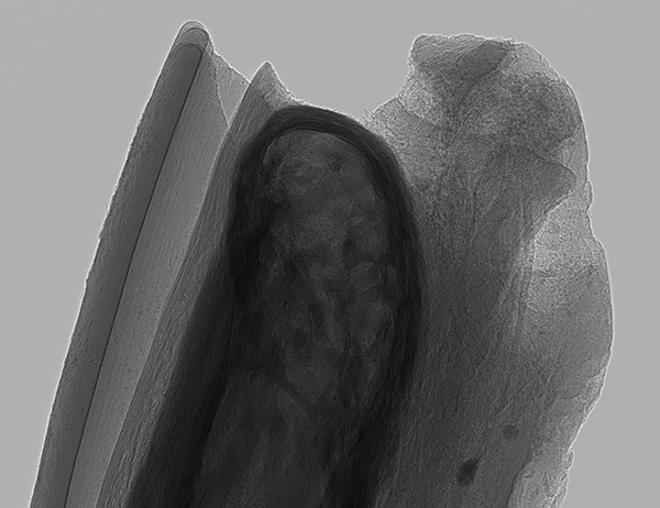
In a recent publication in Radiology, the possibility of using a MetalJet D2+ source for doing non-invasive virtual histology of ancient mummified soft tissue was demonstrated. An overview phase contrast CT scan of the whole mummified human hand shows the overall anatomy whereas a high-resolution scan of the fingertip shows the cellular-size microanatomy at a resolution below 10 µm. The remnants of adipose cells, nerves and blood vessels are visualized
Application of a novel in vivo imaging approach to measure pulmonary vascular responses in mice
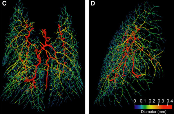
The researchers at Monash University in Australia have successfully been able to transform the in-vivo 4D-CT on pulmonary vasculature in small animals from synchrotron facilities to laboratory with the high brightness microfocus MetalJet X-ray source. Based on this and in conjunction with the dedicated post-image analysis, the quantitative information about the morphologies and dynamics of pulmonary vasculature tree in mice have been extracted. This also indicates that imaging modality can be a complementary methodology added to other in- and ex-vivo studies on murine pulmonary vasculature in small animal models.
M. Preissner, et al., “Application of a novel in vivo imaging approach to measure pulmonary vascular responses in mice”, Physiological Reports (2018).
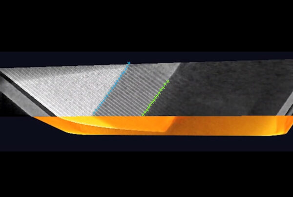
Nucleus-specific X-ray stain for 3D virtual histology
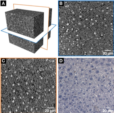
Using a NanoTube, submicron resolution tomography can be obtained by placing the sample very close to the source to generate a large geometric magnification. As an example, at[Field] TU Munich the microanatomy of a mouse liver lobule sample was studied. To obtain contrast, a specific staining method was developed, where the lead-containing stain gives strong attenuation of the X-rays. This enabled three-dimensional visualization of cellular and sub-cellular structures in the sample.
You may find additional imaging application examples on our web.
Other activity
Conferences and Events
At Excillum we are gearing up for a new year of conferences and events, where we will have the opportunity to meet and talk with you! Please, make sure to come by and say “Hi!” any time you see us, whether for a chat and a coffee or to learn more about what you can do with our X-ray sources.
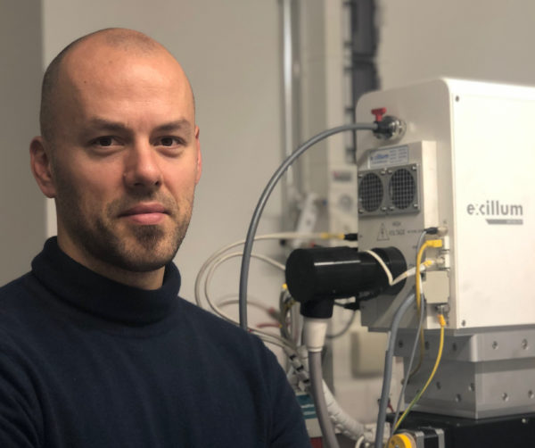
User Story
We met with crystallographer Dr. Alessandro Prescimone in his lab at University of Basel, where we had a very nice talk together with Dubravka Sisak Jung from Dectris.
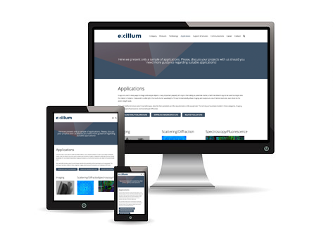
New content on the web
We invite you to check out the new content on the Applications tab on our web site with a whole new section on Imaging.
Career @ Excillum
Excillum is still growing and there are some exciting positions open if you are looking for a challenge. Here is your chance to join an excellent team!

Wishing you all the best and looking forward to seeing you again soon!
The Excillum Team
Published: February 18, 2019