
With the high-brightness, microfocus MetalJet tube, the scientists at KTH demonstrated the ability of laboratory phase-contrast CT in resolving the soft-tissue anatomy and microanatomy in a mummified hand at sub- 10 µm resolution, which is at the cellular-size scale. The imaging methodology used in this study shows its potential in doing non-invasive histology in studies of ancient soft-tissue.
Related Posts
Decision-quality fast CT for battery inspection
Steffen Masuch and Klaus Dröder, TU Braunschweig; and Sophie Gräfnitz, PowerCo.
Ingrid AksnesMarch 26, 2026

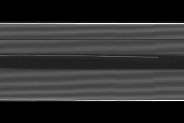
Extremely high-speed X-ray radiography at micrometer resolution to reveal hidden dynamics for failure and root cause analysis of electronics
Julius Hållstedt, Emil Espes, Till Dreier, and Daniel Nilsson, Excillum; Spyridon Gkoumas, DECTRIS Ltd.
Ingrid AksnesDecember 2, 2025
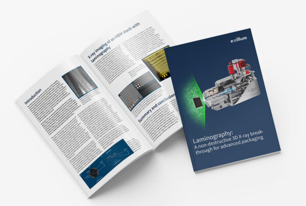
Laminography: A non-destructive 3D X-ray breakthrough for advanced packaging
Till Dreier and Julius Hållstedt, Excillum.
Ingrid AksnesOctober 8, 2025