Small Angle X-ray Scattering (SAXS)
The materials typically studied include polymers, metals, colloids, liquid crystals and biological samples e.g. proteins and RNA/DNA (also referred to as Bio-SAXS). Bio-SAXS has recently gained significant interest in research as well as industry. It has proven to be a valuable technique for the investigation of flexible and disordered proteins and it is complementary to high resolution techniques such as Protein Crystallography and Electron Microscopy.
SAXS measurements can be done on many different types of samples: gels, liquids, solids, etc. Furthermore, little or no sample preparation is required. These two characteristics enable measurements in the native state and in-situ measurements. This gives invaluable insight into the behavior of the analytes in real-life conditions.
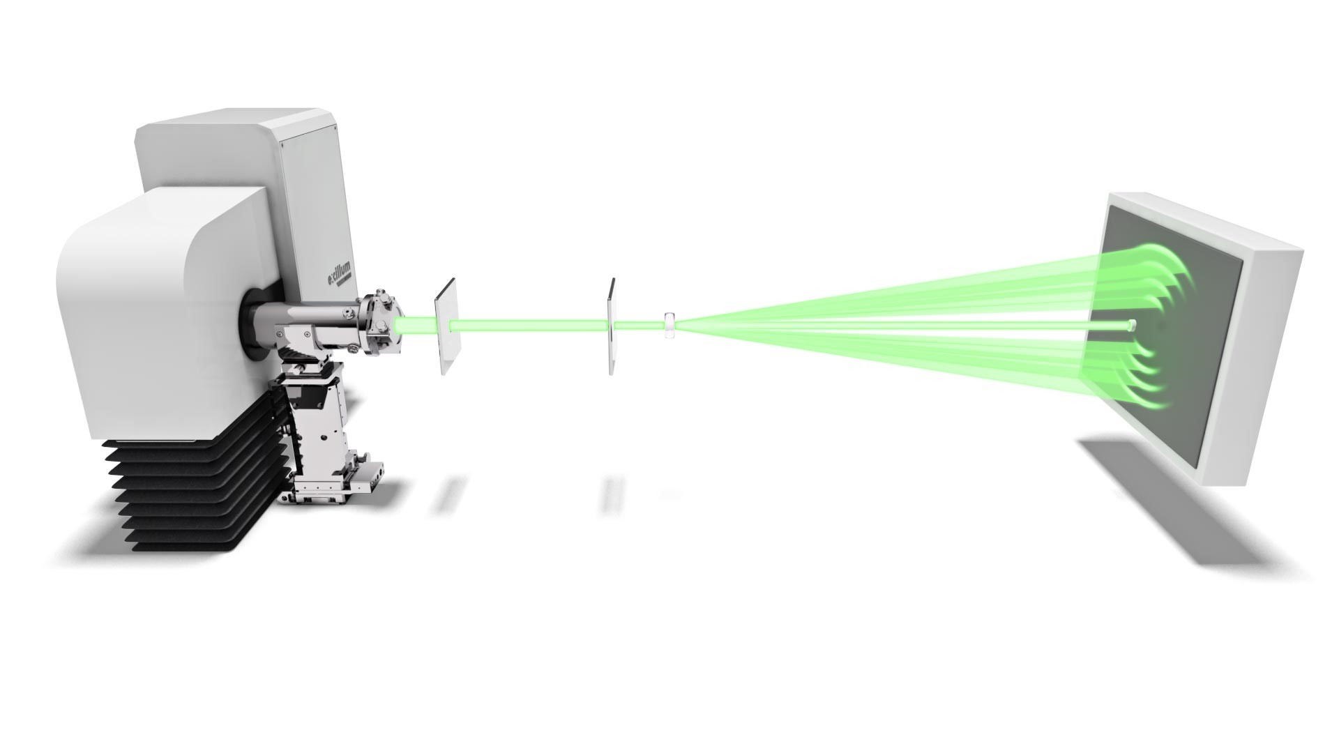
In general, the scattered signal is recorded at diffraction angles below 6° and the sample is positioned at a long distance from the detector. This means that the scattered signal is often weak. Therefore, SAXS measurements benefit greatly from the use of a high brilliance X-ray source such as the Excillum MetalJet. Higher signal means that the weak scattering effects become stronger, more visible and thus more readily studied. Especially time dependent studies (such as the kinetics of particle formation), in-situ measurements (for example under tensile or shear stress) and measurements on weak scatterers like most proteins, benefit hugely from the MetalJet’s high brilliance. Moreover, the use of a high brilliant source reduces the measurement time hereby increasing the amount of measurements that can be done.
Application examples for Small Angle X-ray Scattering (SAXS)
- Biological (Bio-SAXS)
- In-lab SEC-SAXS
- GISAXS/GIWAXS
- In-situ shear
- Fibers
- Metallic nanoparticles
- Polymers
- LDL
Structural characterization of an apoptosis-regulating protein
A MetalJet X-ray source mounted on a Bruker Nanostar was used to help identify the structure of the Bcl-xL protein. This member of the Bcl-2 family plays a role in the regulation of programmed cell death (apoptosis) by governing mitochondrial calcium ion transport. Previous research has shown that Bcl-xL exists in different topological states, mainly as dimers, but structural studies have so far been focused on the monomeric form. Furthermore, two different states of Bcl-xL were known, but the transition between these water-soluble and membrane-bound states remained elusive.
Oligomerization has been shown to be an important step in the process of membrane insertion for other Bcl-2 family members. Therefore, the researchers of Nanyang Technological University, A*STAR, University of Louisville, Rosalind Franklin University of Medicine and Science, and Kyung Hee University set out to resolve the structure of Bcl-xL dimers.
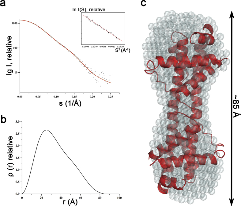
The Bcl-xL dimerization was induced by the addition of a mild detergent (n-Octyl β-D-Maltoside) and the resulting structure was studied by a combination of crystallography, nuclear magnetic resonance (NMR) and Small-Angle X-ray Scattering (SAXS). Protein crystallography showed that the detergent-treated Bcl-xL forms a dimer through three-dimensional domain swapping (3DDS) of helixes α6-α8 between two monomers. The formation of stabile dimers was confirmed by SAXS analysis as indicated by the lack of aggregation which can be seen in the Guinier plot (insert in figure a) and the maximum dimensions (shown in figure b). Furthermore, SAXS measurements yield a low-resolution solution structure of the dimer which fits well with the crystal structure.
The SAXS patterns were acquired for 1 mg/ml and 3mg/ml solutions using a liquid gallium-based MetalJet source with the sample-to-detector distance set to 670 mm using a total acquisition time of 30 minutes.
Structual investigation of protein samples
Small-Angle X-ray Scattering (SAXS) is a prevalent technique in structural biology for the investigation of protein structure and interactions. It gives efficient access to parameters like the radius of gyration, molecular weight, degree of folding, a low-resolution model of the 3D shape of proteins, protein multimers and complexes.
Often, biological samples consist of a complex mixture of structurally different molecules and complexes in dynamic equilibria. SAXS measurements of such a mixture result in an average structure of the species in solution which can be very difficult – if not impossible – to deconvolute. An efficient solution to this problem is offered by the use of Size Exclusion Chromatography (SEC). By fractionating the mixture based on the size of the components, you can measure the SAXS signal of the individual species.
The combination of SEC with SAXS does however put a huge time constraint on the SAXS measurements. Short measurement times are required to prevent the separated samples from reaching a new equilibrium. Short measurements on weakly scattering samples could previously only be done at the synchrotron. With the introduction of the MetalJet X-ray source, this measurement capability is now also available in the laboratory as was demonstrated with the Xenocs BioXolver.
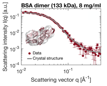
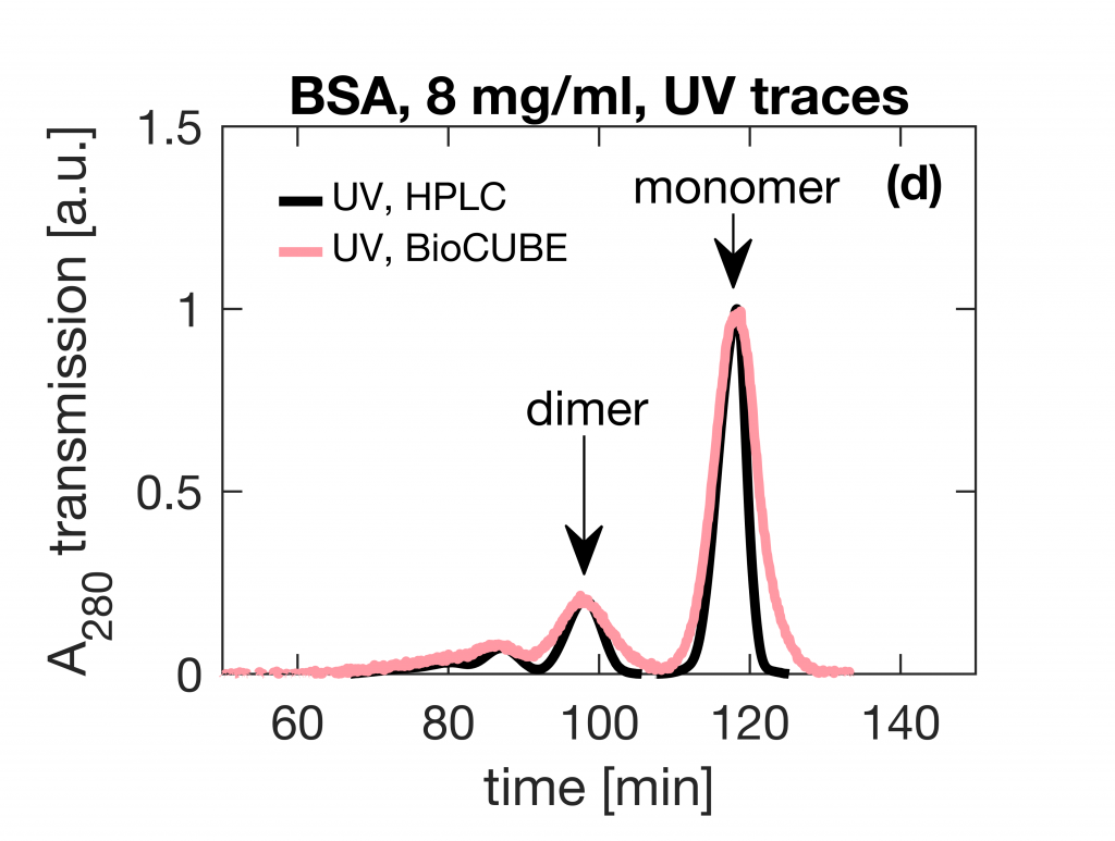
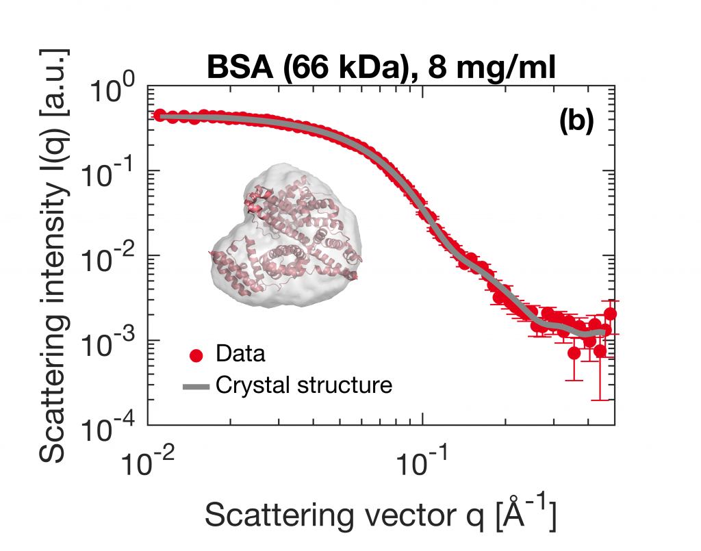
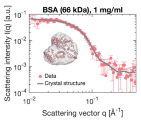
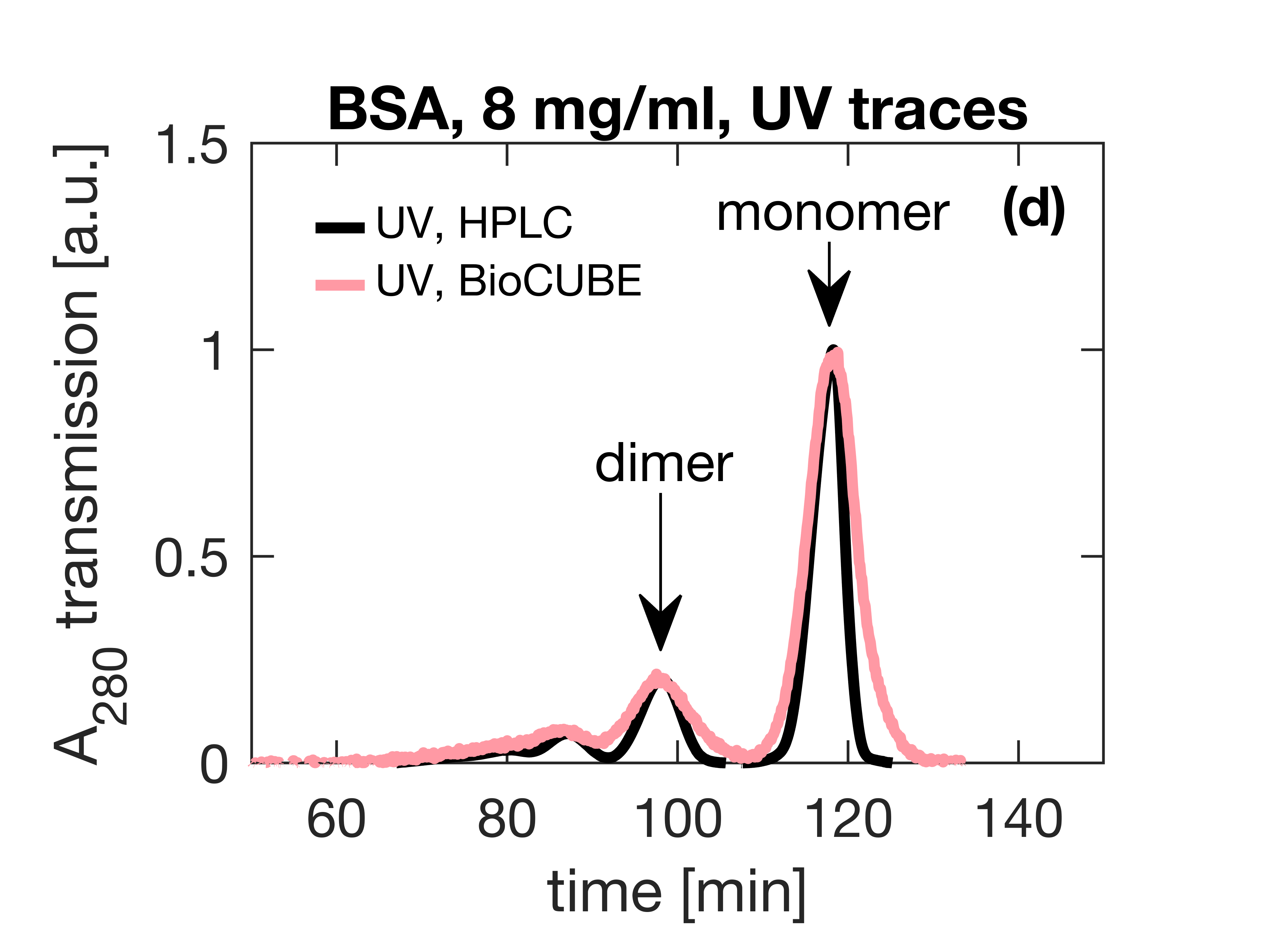
The images below present a case where Bovine Serum Albumin (BSA) was separated into the monomeric and dimeric form. The UV trace shows the separation into monomers and dimers. After separation, these species are measured with SAXS using an exposure time of only 60 seconds. As illustrated below, these short measurements provide sufficient statistics both for concentrations of 8 mg/ml as well as down to 1 mg/ml to establish the most important parameters and obtain a 3D structure.
Application note kindly provided by Application Scientist at Xenocs SAS using a BioXolver equipped with Metaljet X-ray source.
Bucciarelli, S. et al. Size-exclusion chromatography small-angle X-ray scattering (SEC-SAXS) of water soluble proteins on a laboratory instrument, J. Appl. Crystallogr. 2018
High intensity source facilitates in situ-GISAXS measurements on organic solar cells
Grazing-Incidence Small-Angle X-ray Scattering and Wide-Angle X-ray Scattering (GISAXS and GIWAXS) are variations on SAXS and WAXS where, instead of measuring through the sample (transmission mode), the beam reflects from the sample (grazing mode). This change turns the technique from bulk sensitive to surface sensitive. In this type of experiments the X-ray beam hits the sample under a grazing angle (see image below), typically below 0.5˚. GISAXS and GIWAXS are used to give insight into the order and orientation of thin films on a substrate, typical examples include solar cells, long range order of block copolymers, nanoparticles and nanoparticle composites, membranes and lithographic patterning.
Using a high intensity X-ray source such as the MetalJet for these experiments enables in-situ investigation of materials. One example of such an in-situ study is that of Vegso at al. from the institute of physics of the Slovak academy of science. They used a laboratory GISAXS/GIWAXS setup with a MetalJet emitting Ga Kα X-ray radiation to investigate the phase separation of the polymer and the fullerene phase in a bulk heterojunction solar cell.
The size and intermixing of domains of the electron acceptor (polymer) and the electron donor (fullerene) are key determinants of the efficiency with which solar energy is converted to electrical power. The combination of GISAXS and -WAXS give unprecedented insight into the real-time structural evolution of the polymer:fullerene phase separation in terms of polymer crystallization and fullerene agglomeration. GISAXS patterns were collected with 10 second intervals and GIWAXS patterns with 2.5 second intervals. These short exposures were sufficient to follow the kinetics of the bulk heterojunction formation of the spin-coated polymer: fullerene mixture by means of tracking the ratio of assembled material versus non-assembled material (φ) as depicted in the picture to the right, the radius of gyration (Rg) of the fullerene phase and the d100 spacing of the lamellar polymer phase.
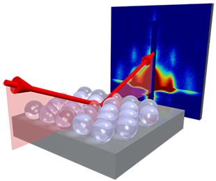
Image credits: www.gisaxs.com Kevin Yager CC BY-SA 3.0
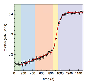
Ratio of assembled versus not assembled polymer-fullerene mixture as calculated from grazing incidence scattering measurements.
Small and Wide-Angle X-ray Scattering of a polymer phase transition under shear
In-situ measurements provide a wealth of information about the sample under investigation. The main asset of in-situ Small and Wide-angle X-ray Scattering (SAXS and WAXS) measurements is that it allows to correlate observations to the underlying structural changes in the material. Measuring a polymer under tensile stress for example, will not only provide parameters like breaking strength, Young’s modulus and strain-hardening properties, but also give insight into what structural changes cause this behavior. Another example is melting behavior of (liquid) crystals, not only will you find the transition temperatures, but WAXS will also reveal which crystal phase is formed. This type of measurement does however put constraints on the measurement time: this needs to be sufficiently short to capture the relevant processes. The use of a high brilliance X-ray source helps to meet these often arduous requirements.
An elegant example of such an in situ study using a MetalJet X-ray source is the work of the Mykhaylyk group at Sheffield university. In this work, they set out to mimic the formation of spider silk with poly(ethylene oxide) (PEO). This is achieved by heating the samples to above the melting temperature, cooling to 25 or 0 ˚C (depending on the exact nature of the sample) and subsequently shearing the aqueous solution to mimic the shear force of extrusion out of silk ducts. Simultaneous collection of Small and Wide-Angle X-ray Scattering (SAXS and WAXS) patterns in situ, show an increase of the scattering intensity as well as the orientation of the sample. These parameters indicate the formation of crystallites and the alignment with respect to the flow direction respectively. The onset of the increase in intensity of both values appears at 60 seconds, which is in agreement with the critical specific work (Wc) calculated from shear-induced polarized light imaging (SIPLI) measurements.
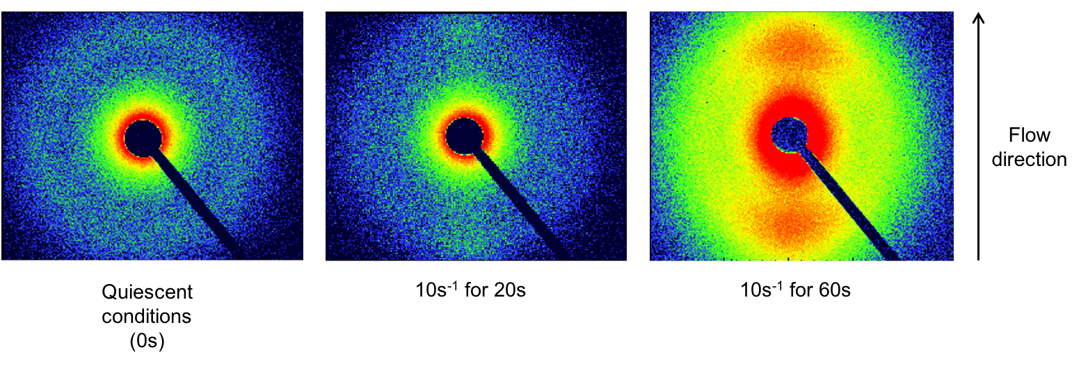
2D Small-Angle X-ray Scattering patterns of PEO solutions sheared at a rate of 10/s for 0, 20 and 60 seconds.
Fibers
Fibers are an important component in many natural and man-made materials ranging from clothes to technologically advanced materials like optical fibers. Many natural fibers, such as for example spider silk, are extremely strong and durable. To mimic the properties of this type of materials in synthetic fibers, a thorough understanding of the fiber structure is required. Small-Angle X-ray Scattering (SAXS) provides an excellent means to obtain information on parameters such as porosity, crystallinity, and the size, shape and dispersity of structural units as well as their orientation. Together, these properties dictate the mechanical properties of the fibers. The technique is also well suited to monitor how these properties change during production and under load and stress.
Especially for the analysis of very thin fibers and for in situ measurements, a high intensity, low divergence X-ray source is advantageous. The high intensity of the MetalJet X-ray source allows short measurement times, even for weakly scattering fibers. For time resolved measurements, such as tensile stretching or the monitoring of an extrusion process, the high intensity is indispensable. The low divergence of the MetalJet ensures good separation between signals, which in turn gives better defined reflections and allows for more accurate integration of the intensities.
The result of a SAXS measurement on a fiber can be seen in the 2D scattering pattern of rat tail tendon above. These thin fibers are a common standard for the calibration of SAXS instruments. Rat tail tendons consist of collagen, which has a primary spacing of about 67 nm and produces a SAXS pattern with equidistant peaks. The above scattering patterns were acquired by application scientists at Bruker AXS using a Nanostar instrument, equipped with an three different X-ray sources: a sealed-tube, solid anode microfocus source, a rotating anode, and an Excillum MetalJet (200W at 70kV). These experiments show that the MetalJet gives an intensity gain of more than 50 times compared to sealed microfocus tubes.
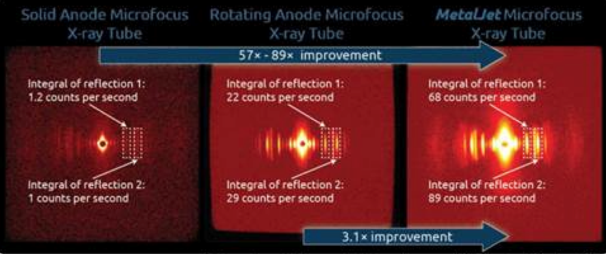
Data courtesy of J. Lange, A. Schwamberger and K. Erlachen of Bruker-AXS.
Metallic nanoparticles
Colloidal metal nanoparticles are a class of materials with myriad applications: drug delivery, medical imaging, catalysis and for antimicrobial purposes, to name just a few. They also show potential as strain gauges. A monolayer of colloidal gold nanoparticles on a flexible substrate offers an interesting alternative to currently used resistive strain gauges. Researchers at the Slovak Academy of Science and STU Centre for Nano-diagnostics show that they provide a fast and linear response is a wide strain range.
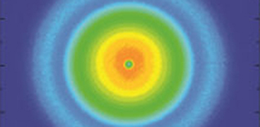
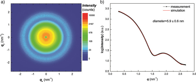
Small-Angle X-ray Scattering (SAXS) gives insight into the electrical response of this new type of gauges by efficiently probing the structural changes of the nanoparticle assemblies induced by stress on the gauge. The use of a high intensity MetalJet X-ray source uniquely enables the researchers to study this system out of equilibrium, during the stretching of the foil. The images below show the 2D scattering pattern (a) of the used gold particles suspended in hexane. Fitting of the radially averaged 1D curve (b) reveals the shape and diameter of the particles.
The MetalJet is available with a gallium-rich (l = 1.34 Å) and an indium-rich (l = 0.51 Å) target. These low wavelengths result in lower absorption, and thereby an increase in the scattering signal. This is especially interesting for high-Z materials such as most metals, which are challenging to measure using a copper source. Furthermore, using these short wavelengths, smaller structures can be probed, air scattering is minimized, which makes in-air measurements more attractive.
These properties, together with the high flux, ensure that short measurements of only 10 seconds, provide sufficient statistics for the analysis of the gold particle monolayers on Mylar foil exposed to uniaxial stress. The in-situ measurements allow the correlation of interparticle distance with the simultaneously measured stress-strain curve, hereby enabling comparison of the macroscopic and microscopic properties of the gauge.
The SAXS measurements were performed in vacuum on a Bruker-AXS Nanostar instrument equipped with a Ga liquid metal-jet X-ray microfocus source.
Polymers
The investigation of polymers is one of the main areas of focus of the Soft Matter Analytical Laboratory (SMALL) at the University of Sheffield. A £2 million infrastructure investment enabled them to purchase a state-of-the-art SAXS instrument with a gallium-based MetalJet X-ray source in 2016. The first publication with their new equipment was a study on the reversible addition–fragmentation chain transfer (RAFT) copolymerization of styrene with N-phenylmaleimide, resulting in the formation of block copolymer nano-objects with a potential application as foam stabilizers.
The researchers at SMALL showed that polymerization-induced self-assembly (PISA) of this copolymer gives different morphologies depending on the ratio between the two polymer blocks. In their article, they present a novel synthesis method using a less toxic cosolvent compared to previous studies: methyl ethyl ketone versus the previously used 1,4-dioxane. Transmission electron microscopy (TEM) revealed vesicular morphologies, similar to what was observed in their previous studies with other PISA formulations. Using Small-Angle X-ray Scattering (SAXS) however, they were able to obtain structural information about the internal structure of these vesicles. This way, they found that in the current system they obtain oligolamellar vesicles, whereas the use of a different stabilizer block results in unilamellar platelet-like particles. Furthermore, SAXS confirms the morphology of the self-assembled copolymers (see figure above) and gives access to statistically relevant information about the size and shape of the self-assembled polymer chains.
The SAXS instrument, equipped with a liquid gallium MetalJet, was the first of its kind to be installed in the UK. The intensity of the X-rays is about a hundred times higher than what could be achieved with the old SAXS instrument. Dr. Oleksandr Mykhaylyk, the facility manager, says that this enables them to analyze their samples in several minutes, rather than the several hours it took with the old instrument.
Using SAXS to gain a deeper understanding of a biomarker for atherosclerosis
Low density lipoprotein (LDL), commonly known as cholesterol, is used as a biomarker for atherosclerosis (the narrowing of arteries due to the formations of lesions). Over the past years however, it has become clear that just the amount of LDL found in a patient’s blood is not an accurate predictor of the risk for atherosclerosis. Low density lipoprotein is a mixture of lipids, triglycerides, and sterols of which the structure and composition is different across individuals. Insight into the different LDL components and their structure is crucial for understanding and prevention of atherosclerosis.
Complex biomolecular assemblies are some of the most challenging systems to characterize in terms of structure and function by means of biophysical characterization techniques. Crystal structures are difficult to obtain due to the difficulty to crystallize the heterogeneous samples, and Cryo-TEM (cryogenic Transmission Electron Microscopy) requires freezing the species into a fixed conformation. By contrast Small-Angle X-ray Scattering (SAXS) measurements can be done under (near) physiological conditions and offers the possibility of in-situ studies such as under influence of temperature changes, oxidation and even the addition of possible anti-atherosclerotic drugs. Traditionally, this type of measurements could only be done at synchrotron facilities, but the introduction of high brilliance sources means that this can now be done in-house as demonstrated by Maric et al. in a collaboration between Malmö university and Aarhus university.
The researchers developed a model describing LDL particles based on temperature dependent SAXS (see picture) and cryo-TEM data. This model successfully describes the size and shape as well as the lamellar internal arrangement of LDL particles and the transition temperature at which the internal structure disappears. Previous studies have shown that the temperature induced transition of the internal structure of the particles is patient dependent and that the ordered arrangement of the core lipids has an effect on the resistance to oxidation, which is known to worsen atherosclerosis. The model was successfully used to describe LDL particles obtained from human blood samples and is expected to enable comparisons between different risk groups in the future.
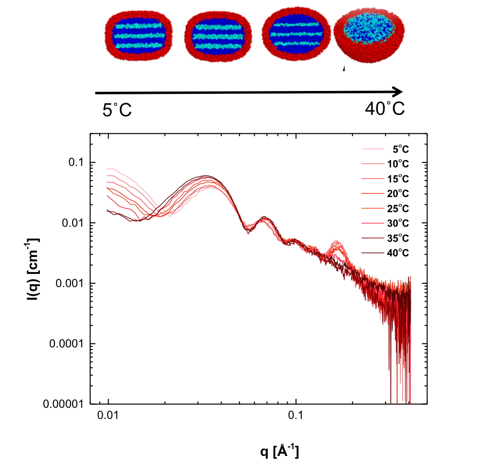
Radially averaged SAXS patterns of LDL, collected at temperatures between 5 and 40˚C.
Related user stories
Experiences with the Excillum X-ray sources
Learn about some of our customers' experiences with our state-of-the-art X-ray sources.