X-ray spectroscopy and fluorescence
X-ray spectroscopy and fluorescence is used in many fields, such as material science, biology, forensics, environmental science and industrial applications.
The object can be characterized either by how its absorption and scattering change with respect to the energy of the incident X-ray beam or by the energies of the photons and electrons that are emitted from the object. What can be measured varies largely over the range of spectroscopic methods, as does the requirements on the equipment. When elemental contrast and spatial resolution are to be combined, the incident X–ray radiation is commonly focused into a small spot or beam at the object. This requires both an X-ray source with high brightness and X-ray optics designated for the specific task.
Application examples for X-ray spectroscopy and fluorescence
High Definition Maia Mapper laboratory-scale XRF imaging system based on the MetalJet source
The Maia detector array was initially developed for high-resolution X-ray fluorescence imaging at synchrotrons. Today, thanks to the high brightness, tight emission regulation and well-fitted spectrum of the Excillum MetalJet X-ray source, this technique is possible to use also at a compact laboratory setup.
The MetalJet source was equipped with a polycapillary lens to give a 32 µm focus determining the spatial resolution. Using translation stages with long range, the sample can be scanned over large area in high resolution. The exposure time is kept low thanks to the high brightness MetalJet X-ray source and the high flux gain in the optics.
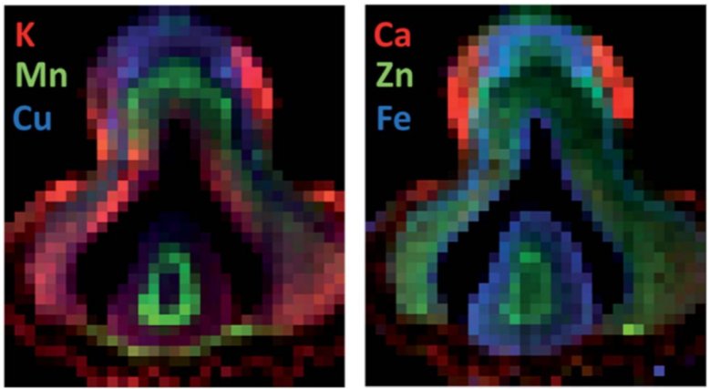
The example of a drill core sample shows the potential of the method to identify a multitude of elements simultaneously, in this case calcium, iron, manganese, rubidium, strontium and gold.
Read user story
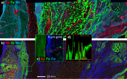
A drill core from the Jundee gold mine imaged with the Maia Mapper showing the combination of large field of view (231×46 mm2) and high resolution (30 µm pixel size) within decent acquisition times (6.0 ms dwell). The RGB images are shown here as Ca-Fe-Mn (a) and Rb-Sr-Mn (b), the inset from Au-Fe-Ca image highlights the details within the region with a rare grain and the spectrum thereupon.
C.G. Ryan, et al., “Maia Mapper: high definition XRF imaging in the lab”, J. Instrum. (2018).
High-spatial-resolution x-ray fluorescence tomography with spectrally matched nanoparticles
By aligning a multilayer Montel mirror to a MetalJet source, a semi-monochromatic, 100 µm narrow beam with low divergence can be created. This was implemented at the Royal Institute of Technology in Stockholm, in order to use the beam as excitation for x-ray fluorescence imaging of mice. As contrast agent, the mice are injected with molybdenum nanoparticles that are passively targeted to tumors but also show up in other organs.
The setup contains one detector behind the sample to measure the transmission and one detector on the side to measure the fluorescence, and the object placed on a motion stage. Translation and rotation of the object allows tomography by a point-by-point acquisition, which is followed by an iterative reconstruction to obtain the quantitative three-dimensional distribution of nanoparticles in the mouse. The experiments have short acquisition time, low radiation dose and low nanoparticle dose which make in vivo experiments possible.
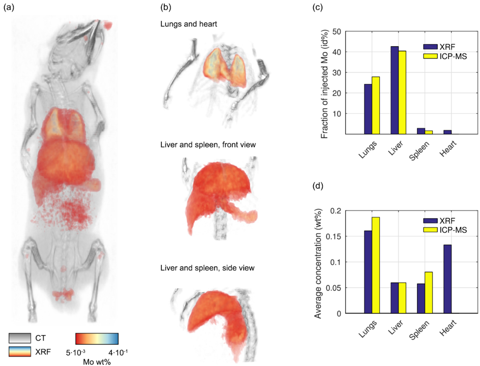
Overlay of the 3D-reconstructions of the conventional CT and the x-ray fluorescence signal, acquired simultaneously. The images show an ex vivo mouse and the nanoparticle concentration in its organs. The quantitative results provided in the method agree well with measurements in ICP-MS.
C. Larsson, et al., “High-spatial-resolution x-ray fluorescence tomography with spectrally matched nanoparticles”, Phys. Med. Biol. (2018).
Hard X-ray photoelectron spectroscopy (HAXPES)
Hard X-ray photoelectron spectroscopy (HAXPES) measurements offers the possibility to investigate material properties by analyzing the photoelectrons emitted from the sample when exposed to hard X-rays. The depth below the sample surface and the number of atomic elements that can be analyzed depends on the X-ray energy. Higher energies allow analysis of bulk properties of various materials, buried interfaces and access to deep core levels.
Based on the Excillum MetalJet technology, HAXPES-Lab by Scienta Omicron, is the first laboratory instrument for photoelectron spectroscopy with an excitation energy of 9.25 keV. Previously, such high excitation energies have only been possible at synchrotrons, since the small interaction cross-sections are these energies put a high requirement on the incident flux.
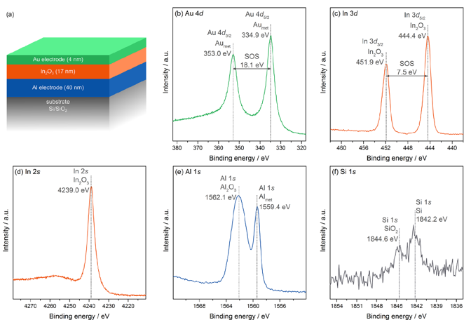
(a) Single-oxide structure for transistor and diode fabrication and (b-f) core-level measurements by the HAXPES-Lab instrument.
A. Regoutz, et al., “A novel laboratory-based hard X-ray photoelectron spectroscopy system”, Rev. Sci. Instrum. (2018).
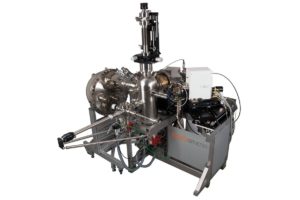
HAXPES-Lab setup based on a MetalJet source
Confocal micro-X-ray fluorescence spectroscopy with a liquid metal jet source
A setup with focusing optics for both the excitation and detection, in a confocal arrangement, allows three-dimensional spectroscopic imaging. This type of experiments is traditionally performed on synchrotrons, since the combined yield from the fluorescence and through the optics become very low. When performed at conventional solid-anode sources, scan times for typical samples tend to be a few days or even weeks.
Researchers at the Technical University of Berlin, Germany, worked with a setup for confocal micro–X-ray fluorescence spectroscopy. The device is based on an Excillum MetalJet source equipped with polycapillary optics. The Gallium emission lines at 9.25 and 10.27 keV are focused into a 31 µm focus which is used to excite x-ray fluorescence in the sample. The fluorescence is collected by a second polycapillary lens and detected by a spectrometer. Examples show that the device provides both high spatial resolution and high contrast sensitivity for a range of elements.
Confocal x-ray fluorescence images of a virtual slice in a millet seed. The colors display the concentrations of the elements. The voxel size is 33x37x37 µm.
L. Bauer, et al., “Confocal micro-X-ray fluorescence spectroscopy with a liquid metal jet source”, J. Anal. At. Spectrom. (2018).
Unless otherwise stated, pictures and content is published under license for CC-BY (https://creativecommons.org/licenses/by/4.0/).
Related user stories
Experiences with the Excillum X-ray sources
Learn about some of our customers’ experiences with our state-of-the-art X-ray sources.