Pushing the limits in X-ray fluorescence
In mineral exploration, locating traces of rare and precious metals is like finding the proverbial needle in a haystack. From a drill core sample up to three kilometers in length, the researchers at CSIRO in Australia are tasked with locating, mapping and characterizing extremely low concentrations of metal particles, often no larger than a few specks of dust.
Interviewees
Chris Ryan, Senior Principal Research Scientist
Institute
CSIRO Advanced Resource Characterization Facility, Australia
Method
High-definition X-ray fluorescence imaging
Application
Mineral resource characterization
X-ray sources
Excillum MetalJet D2+
You’re approaching an order of magnitude more out of the MetalJet and an order of magnitude more out of Maia. Put those together and you’re really getting some high throughput.
Chris Ryan
Imaging minerals from drill core to atomic scales
CSIRO’s advanced characterization process operates on a wide range of spatial scales to help the Australian minerals industry apply research outcomes to their ore deposits. Ore deposits produce hundreds of kilometers of drill core a year. The core is analyzed in the lab or onsite by high-speed X-ray fluorescence (XRF) and hyperspectral linear core scanning instruments provided by mining equipment and technology services companies. Research delivers high resolution images and datasets from small subsamples using synchrotron radiation, scanning and transmission electron microscopy, nano-scale ion probe analysis and atom probe tomography. CSIRO is linking these scales by imaging fifty-centimeter lengths of core, using a laboratory micro-XRF system called Maia Mapper based on an Excillum MetalJet X-ray source. Bridging this gap means that the results of the micro-to-atomic scale studies can be scaled up because we can place this detail in the context of the drill-core samples.
Within the analysis workflow, stretching from kilometers to a few atoms, the MetalJet was chosen for its ability to fill an important spatial imaging gap. “We already have an advanced detector array called Maia for use at the synchrotron,” explains Chris Ryan, Senior Principal Research Scientist, “so we started to discuss whether it could be used in a lab-based setting for handling intermediate spatial scales, prior to synchrotron analysis.”
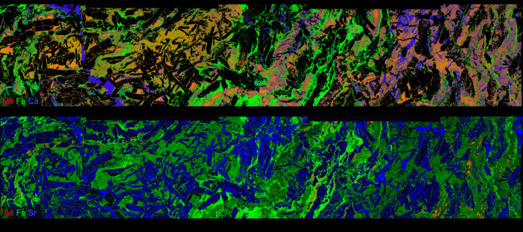
12 M pixel RGB false-color images of a drill core segment (236 x 46 mm2) from the Abra lead-silver-copper-gold deposit in Western Australia (7867 x 1533 pixels, each 30 μm at 5.3 ms dwell time) acquired using Maia Mapper and an Excillum MetalJet D2+ at 200 W and 70 kV into an effective 20 μm source size.
Two pioneering technologies
The main criterion for the new X-ray source was maximum brightness, in order to produce the highest-intensity focused X-ray beam using a polycapillary lens. “This suggests that you want a focus of twenty microns or less at the source, with maximum brightness,” Chris explains. “We were trying to go for a system that would push things to the limit so we would get maximum pixel throughput at good sensitivity and make full use of the capability of our Maia detector array, which can handle many millions of counts per second. You’re approaching an order of magnitude more out of the MetalJet and an order of magnitude more out of Maia. Put those together and you’re really getting some high throughput.”
Because we’re doing imaging where we scan a sample through the focused beam spot, any fluctuation in the intensity of the beam will be visible as an artifact in the image. So we’re very sensitive to this, and our imaging has proved to be just brilliant. It’s clearly the best quality XRF imaging with any source.
In addition to brightness, stability was also a key concern when building the new system. The reliability of the source, the stability of the flux and the steadiness of the source spot were all essential for ensuring a reliable focused flux. “Excillum did some very nice tests of this for us, so we could satisfy ourselves that it was behaving as we like,” says Chris. “Because we’re doing imaging where we scan a sample through the focused beam spot, any fluctuation in the intensity of the beam will be visible as an artifact in the image. So we’re very sensitive to this, and our imaging has proved to be just brilliant. It’s clearly the best quality XRF imaging with any source.”
The institute now has MetalJet sources in two labs, with both systems running up to 24/7 with the expected stability. The first source installed has been running for more than two years now with a beam intensity fluctuation of less than 0.1 percent. “In other words, it’s hard to even detect, and we don’t see any fluctuation at all in our images,” says Chris.
Expanding the search space
Within the field of exploration, ore deposits are often surrounded by halos, or footprints, of particular elements that you might not think are related to the commodity you are looking for. While thallium halos around some of Australia’s largest zinc deposits have long been known about, he explains, understanding which minerals the thallium occurs in within the rocks and how these minerals relate to the zinc is a significant advance that has come from the Maia Mapper. We can also hunt out particles of precious metals like gold or platinum. Ores can contain as little as a few parts per million of these metals and we need to understand what elements are associated with the precious metals to find more and also to extract them from the rocks. With only a handful of particles in a piece of core this is a real challenge for conventional instrumentation. “We can handle a half-meter length by eighty millimeters in diameter, for a large drill core, and image that whole area with a resolution of 30 μm” says Chris. “This means we end up with twenty, thirty or up to fifty million pixels of image data. It enables us to do things that were very difficult before, where we can find objects like precious metal particles that are distributed quite rarely over large areas. Now we can sample enough of these rare particles to get a clearer picture of the mineral associations and features they’re associated with, such as generation of vein in the rock. This is very difficult to do if you just see one or two of these with normal microanalysis, which is only spanning a millimeter or so. This intermediate spatial scale helps us see the bigger picture.”
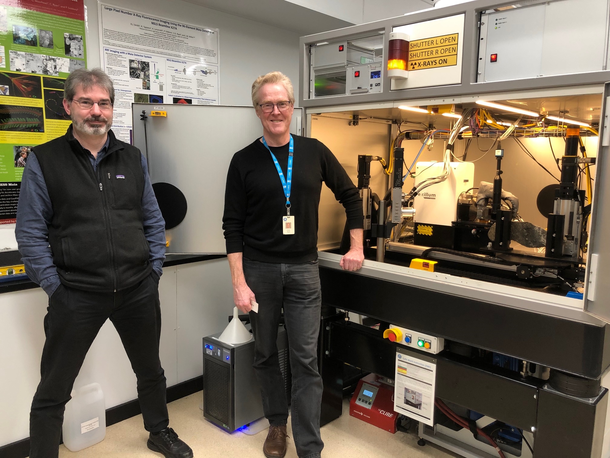
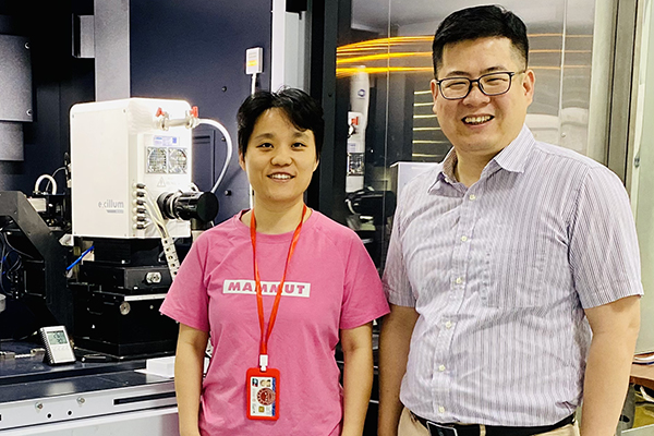
Robin Kirkham (Lead Engineer) and Chris Ryan (Senior Principal Research Scientist) in front of the Maia Mapper and Excillum MetalJet setup.
Beyond the lateral coverage, Chris is also impressed by his system’s ability to maintain a level of sensitivity that is needed to identify trace elements at greater depth. “The system has the sensitivity that, even in a 30 μm pixel, we can still see a gold particle that’s only a couple of microns in diameter,” he says. “And since these are such high-energy X-rays, those particles can be tens of microns, or even a hundred microns, below the surface of the sample we’re looking at. Hence, we’ve increased the search space laterally with these large areas at 30 μm resolution, and increased the search volume with depth as well, which greatly increases the chance of finding these rare particles. It’s been good for platinum group minerals, gold, rare minerals in meteorites, and samples from the earth’s mantle that are carried to the surface, for example.”
Handling all of this data, of course, is no small challenge itself. This is why the researchers at CSIRO have spent years developing the data processing capabilities required for high-throughput analysis of such data-rich full-spectral images. “One of the things we’re most proud of,” says Chris, “is combining the large detector array technology developed at Brookhaven National Labs with the real-time processing capabilities that have been central to the work of the CSIRO group for many years. We do the spectral deconvolution in real time. So even if the image has tens of millions of pixels in it, and we’re analyzing each one at a time at two to ten milliseconds per pixel, we actually have the unfolded element concentrations available in real time.”
High-throughput industrial analysis
These combined capabilities, among others, make the setup especially well-suited to potential lab-based industrial applications in association with synchrotrons, says Chris. “We’ve been suggesting to the synchrotrons that a lab-based Maia Mapper and MetalJet setup could be a very efficient way to handle these intermediate spatial scales. Then they can feed only the subsamples or a smaller area onto the synchrotron beam lines, which are much more expensive to run, and then a lot of the industrial applications would be handled in a lab on the side. My argument is that a lot of industrial users are often using macroscopic objects – they’re worried about a weld in a steel product or something – and the scale that we map over hundreds of millimeters really suits the scale of their application. And then if particular defects are identified, you could then take subsamples over to the synchrotron to see things in finer detail. So, I would suggest that it provides an efficient feeder of industrial applications into the synchrotron.”
We’ve been suggesting to the synchrotrons that a lab-based Maia Mapper and MetalJet setup could be a very efficient way to handle these intermediate spatial scales.
A powerful partnership
As the CSIRO group continues to open new possibilities with their cutting-edge technologies, Chris is pleased to have the support of Excillum in exploring and refining these new methods. “The experience of working with Excillum has been really good,” he says. “They’re with us for the journey, if you like. And I think that’s very important when you’re trying to push something to the limit. Like the stability issue, which might not be an issue for many applications, but it’s critical for imaging. So that 0.1% fluctuation: that’s not a parameter that X-ray source manufacturers just quote in a table. You have to get them thinking about and measuring these performance figures for the first time, and Excillum was happy to do that. So not only was MetalJet the brightest source, but we could tick off our concerns about these uncertain parameters that no one talks about. They’re not only understanding of a research environment, and scientists asking tricky questions where they have to come up with an answer, but they’re also producing a product that has the reliability. “
The experience of working with Excillum has been really good. They’re with us for the journey, if you like. And I think that’s very important when you’re trying to push something to the limit.
The development and construction of Maia Mapper is supported by the Science and Industry Endowment Fund (SIEF). The Advanced Resource Characterisation Facility (ARCF) is supported by SIEF. The Abra samples were collected as part of the Capricorn Distal Footprints Project supported by SIEF.