Biomedical research
X-rays can be used on both ex-vivo and in-vivo samples to gain a clearer understanding of biological tissues. However, one of the core challenges in this type of X-ray imaging is to achieve the ideal combination of contrast, resolution and speed. In particular, low-contrast samples such as soft biological tissues demand X-ray imaging systems that achieve the highest possible resolution within the shortest possible exposure time, which in turn creates images with more detailed information for more rapid diagnosis. Whether the research involves phase-contrast tomography for dynamic imaging of live mice, or high-resolution PBI tomography of ex-vivo tissue or high-resolution nanoCT Excillum X-ray sources are proven to excel in a wide range of biomedical imaging applications.

Application examples
High resolution propagation-based imaging system for in-vivo dynamic computed tomography of lungs in small animals
By operating a MetalJet D2+ at 250 W and 15 µm spot size, it has been shown that phase-contrast tomography can be used for dynamic imaging of live mice. In research work done in Australia, time-resolved computed tomography was performed to image the ventilation in the lungs of a mouse. A flat-panel detector acquired projections with only 18 ms exposure time, allowing a full tomography in 32 s. These very short exposure time and controlled breathing, allowed small airways down to 55-60 µm in diameter to be imaged dynamically. This high quality dynamic imaging of lungs enables determination of the lung function, even down on a regional level. Furthermore, high quality dynamic CT potatially has many other applications.
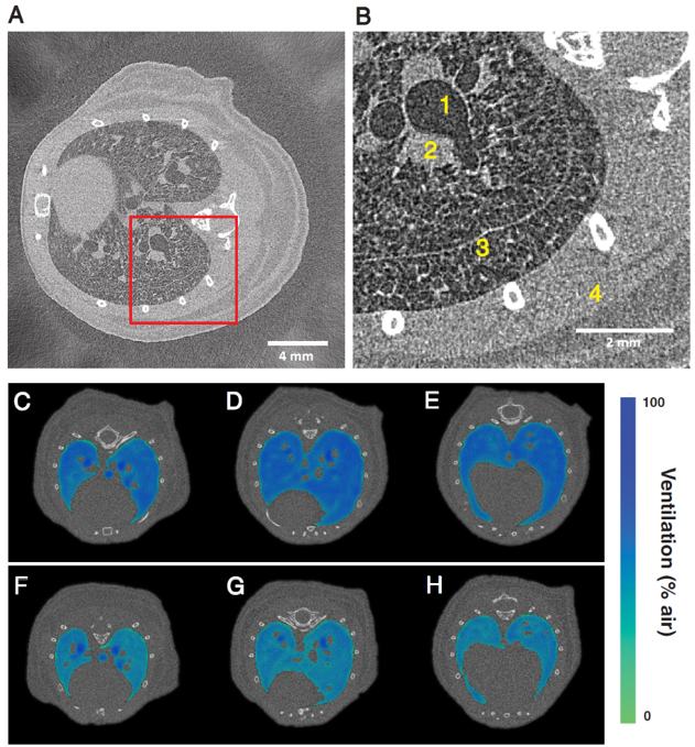
Time-resolved computed tomography of a live mouse (A). Close-up region (B) shows the anatomical features. The method demonstrates the differences in volume of air in the lungs after 0 hours mechanical ventilation (C)-(E), and after 2 hours (F)-(H). Image reprinted from M. Preissner et al., “High resolution propagation-based imaging system for in vivo dynamic computed tomography of lungs in small animals”, Phys. Med. Biol. (2018).
X-ray video of a breathing mouse acquired using an Excillum MetalJet D2+ X-ray source. The frame rate is 30 Hz and effective pixel size 19 µm. Video from M. Preissner et al. “Application of a novel in vivo imaging approach to measure pulmonary vascular responses in mice”, Physiol. Rep.(2018).
High resolution X-ray PBI lung tomography on whole mice at laboratory scale
The scientists at University of Göttingen demonstrated the high resolution PBI tomography on whole mice based on using a MetalJet source. With the optimized spectrum they demonstrated the applicability of PBI in local tomography for small animal imaging of lungs, in the presence of highly absorbing surrounding tissue at µm- level resolution. The result indicated the application of this imaging method at cellular level for even larger biological samples.
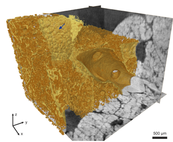
3-D rendering of the PBI tomogram of the soft tissue in the thorax.
- Exposure time = 10 s/ radiography
- voxel size = 5 µm
- Tube voltage = 70 kV, tube power= 100 W
- Timepix photon counting detector
M. Krenkel, et al., “Propagation-based phase-contrast tomography for high-resolution lung imaging with laboratory source“, AIP Adv. (2016).
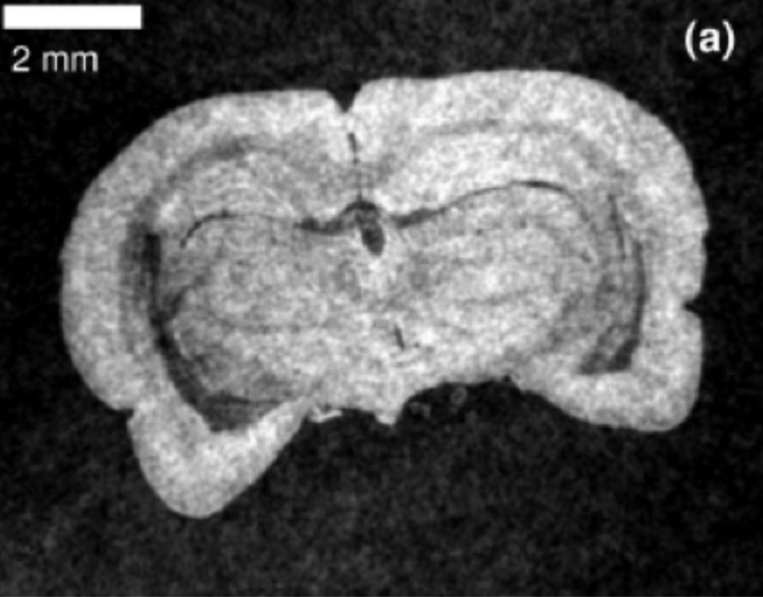
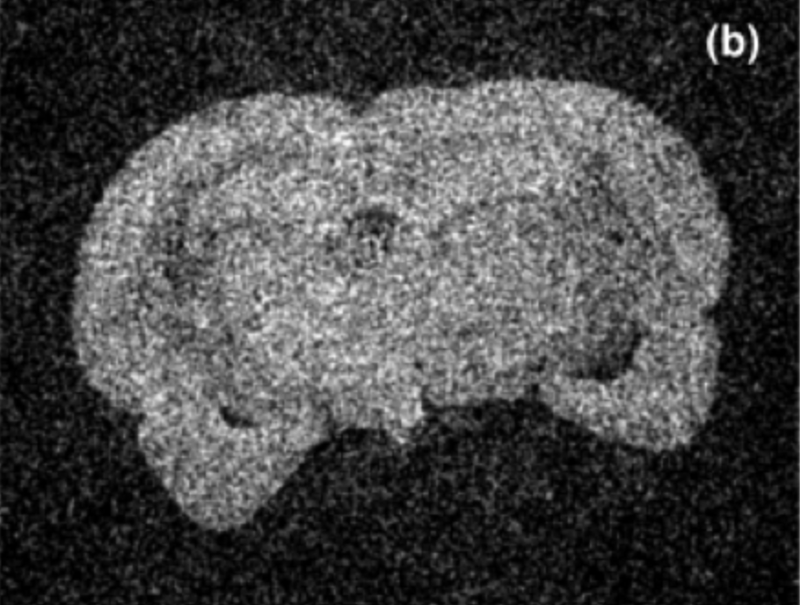
Cellular and sub-cellular resolution X-ray PBI tomography on mouse brain cytoarchitecture
An another example from the research group in Göttingen more recently, the mouse brain cytoarchitecture was investigated at sub-celluar level resolution by PBI tomography. Together with suitable phase-retrieval algorithm and novel tissue prepartation, this study highlights that this is a non-destructive imaging method in the studies of brain architecture in mammals.

PBI tomography slice along transverse (a) and another along longitundnal (b) directions; automatic volume rendering of the reconstructed volume (c) and the zoomed-in cellular segmentation (d). The molecular layer (ML), granular layer (GL), white matter (WM) and Purkinje cell layer (PCL) of the cerebellar vermis at cellular resolution.
- Exposure time = 50 s/ radiography
- Voxel size = 47 µm
- Tube voltage = 40 kV, tube power= 50 W
- Lens-coupled scintillator-based camera XSight
M. Töpperwien, et al., “Three-dimensional mouse brain cytoarchitecture revealed by laboratory-based x-ray phasecontrast tomography“, Sci. Rep. (2017).
X-ray GBI microscope with MetalJet source
The joint research by scientists from KTH and ETH/PSI proved the advantage of using a MetalJet source in GBI, which significantly improved flux and visibility compared to a conventional microfocus tube. Moreover, a preliminary exploration of its biomedical application has been shown by tomography on a rat.
A comparison between phase-contrast (a) and attenuation-contrast (b) slices from GBI tomography based on a MetalJet source. The sample was a rat brain scanned in a liquid paraffin bath.
- Exposure time = 1 min/ stepping image
- Voxel size = 20 µm
- Tube voltage = 50 kV, tube power= 40 W
- G1 is designed for π-phase shift at 25 keV, period= 4.12 µm
- CCD camera with Gadox scintillator
Reproduced from T. Thüring, et al., “X-ray grating interferometry with a liquid-metal-jet source“, Appl. Phys. Lett. (2013) with the permission of AIP Publishing.
High-resolution zebrafish muscle imaging with X-ray PBI tomography
By using a MetalJet tube, PBI tomography of an unstained whole zebrafish was performed. This shows the capability of capturing the low contrast sub-5 µm details with suffciently high contrast. The methodology paves the path for the non-invasive whole-body imaging at sub-cellular resolution for soft tissue research and small animal models, thus facilitate the deep understanding of muscular disease and assessing interventions.
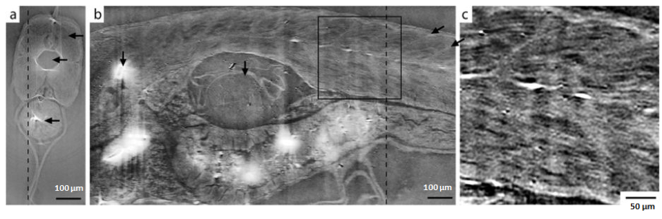
The PBI tomographic axial (a) and sagittal (b) slices and the zoom-in ROI (c) as the black box in (b).
- Exposure time = 115 s/projection
- Voxel size = 0.733 µm
- Tube voltage = 40 kV, tube power = 24 W
- FDI- VHR CCD camera with Gadox scintillator
W. Vågberg, et al., “X-ray phase-contrast tomography for high-spatial-resolution zebrafish muscle imaging“, Sci. Rep. 5. 16625 (2015).
High-resolution nanoCT
A NanoCT device comprising of an Excillum NanoTube and a photon counting detector provides the ability of tomography with very high resolution. At the Technical University of Munich, Germany, the device has achieved a state-of-art ~100 nm spatial resolution and capability of investigating phase-contrast imaging.
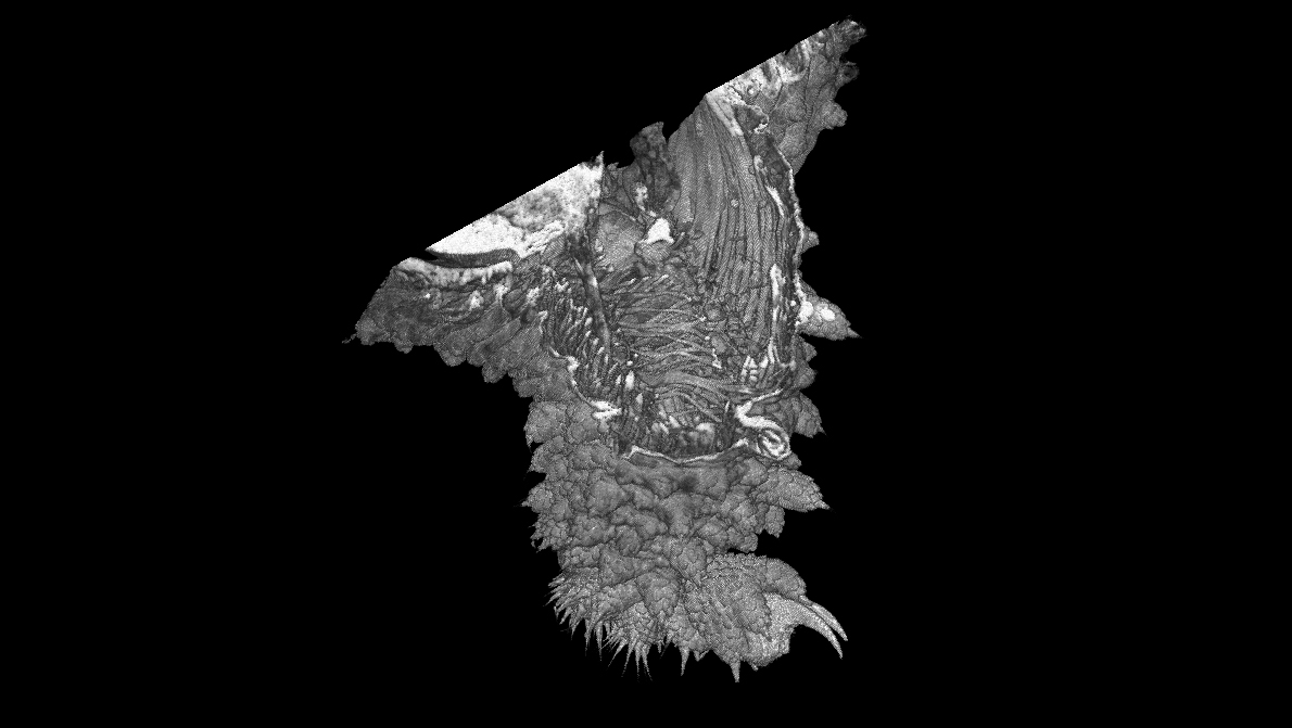
Nano-CT video and image of the limb of a velvet worm (Onychophora), 0.4 mm long. The surface morphology can be visualized with an image quality similar to scanning electron microscopy, and simultaneously the visualization of internal musculature at a resolution higher than confocal laser scanning microscopy.
Related X-ray method
Related user stories
Experiences with the Excillum X-ray sources
Learn about some of our customers' experiences with our state-of-the-art X-ray sources.