Phase-contrast imaging
Phase-contrast imaging requires special techniques to be detected but can be very beneficial. For materials that have low absorption, such as soft biological matter, polymers and many other organic compounds, the phase can give more than 1000 times stronger contrast than absorption. Without going into the detailed physics behind phase contrast, we can still understand the methods that are used to exploit it.
To make phase-contrast imaging, the phase shift induced by the object must be measured. The unfortunate catch is that X-ray detectors don’t measure phase, but only intensity. The phase shift is instead converted to intensity variations, that can be detected. There are several methods to do this, which are all good in different ways.
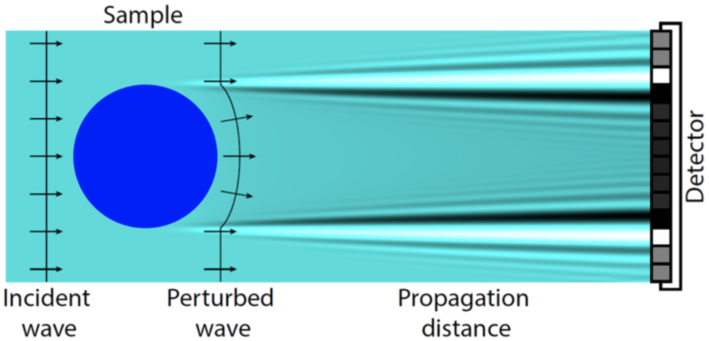
Simple example of how the phase shift through an object perturbs the wavefront. The wavefront cannot be directly measured but is here converted to intensity variations at a distance. The contrast arises from both absorption and phase. The edge enhancements introduced by the phase shift is apparent along all contours.
Compared to absorption-contrast imaging, the requirements on the equipment are in general higher for phase-contrast imaging. To be able to obtain contrast, an X-ray source with high transverse coherence is needed. This means that the source either has to have a small emission spot or has to be moved far away from the object. Both options are traditionally related to very low flux, which leads to very long exposure time. To keep the exposure time short, an X-ray source with a small emission spot that can still maintain a high flux is very beneficial. Another commonly used metric is the source brightness. The source brightness is a measure that includes both coherence and flux and is important to have high in many cases!
Phase-contrast imaging was originally developed for biomedical applications, where it is very beneficial for imaging of soft tissues. Recently, it has seen a growing interest also in materials science, engineering and industrial non-destructive testing.
Propagation-Based Phase-Contrast Imaging (PBI)
PBI has the simplest measurement geometry, similar to conventional attenuation-contrast imaging and requires no optical elements. The only difference is that free-space distances are kept between the object, sample and detector. This leads to that the phase can generate intensity variations, visible as edge enhancements. The image represents a mixing of the absorption-contrast signal and the phase-contrast signal. Implementing PBI in a compact laboratory-scale setup puts high requirements on the spatial coherence of the source, detector resolution and long distance between the sample and the detector. Both the MetalJet and the NanoTube sources meet the requirements for doing laboratory-based high-resolution PBI. Furthermore, the high brightness helps to keep the exposure time down, while still enabling high resolution.
To obtain an image that represents the object thickness, the phase-contrast image must be treated with a process called phase retrieval. There are many methods for phase retrieval, most of them based on convolutions. If using a single image, assumptions on the object must be made, such as assuming a single material object. With multiple images acquired at different propagation distances, this assumption can be circumvented. On the other hand, multiple images introduce a higher complexity in the experiments.
Application examples for Propagation-Based Phase-Contrast Imaging (PBI)
- In-vivo dynamic computed tomography
- Muscle imaging
- X-ray PBI lung tomography
- X-ray PBI brain tomography
High resolution propagation-based imaging system for in-vivo dynamic computed tomography of lungs in small animals
By operating a MetalJet D2+ at 250 W and 15 µm spot size, it has been shown that phase-contrast tomography can be used for dynamic imaging of live mice. In research work done in Australia, time-resolved computed tomography was performed to image the ventilation in the lungs of a mouse. A flat-panel detector acquired projections with only 18 ms exposure time, allowing a full tomography in 32 s. These very short exposure time and controlled breathing, allowed small airways down to 55-60 µm in diameter to be imaged dynamically. This high quality dynamic imaging of lungs enables determination of the lung function, even down on a regional level. Furthermore, high quality dynamic CT potatially has many other applications.
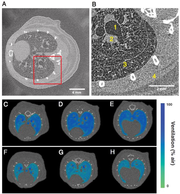
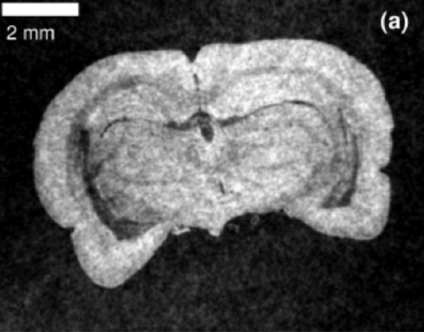
Time-resolved computed tomography of a live mouse (A). Close-up region (B) shows the anatomical features. The method demonstrates the differences in volume of air in the lungs after 0 hours mechanical ventilation (C)-(E), and after 2 hours (F)-(H). Image reprinted from M. Preissner et al., “High resolution propagation-based imaging system for in vivo dynamic computed tomography of lungs in small animals”, Phys. Med. Biol. (2018).
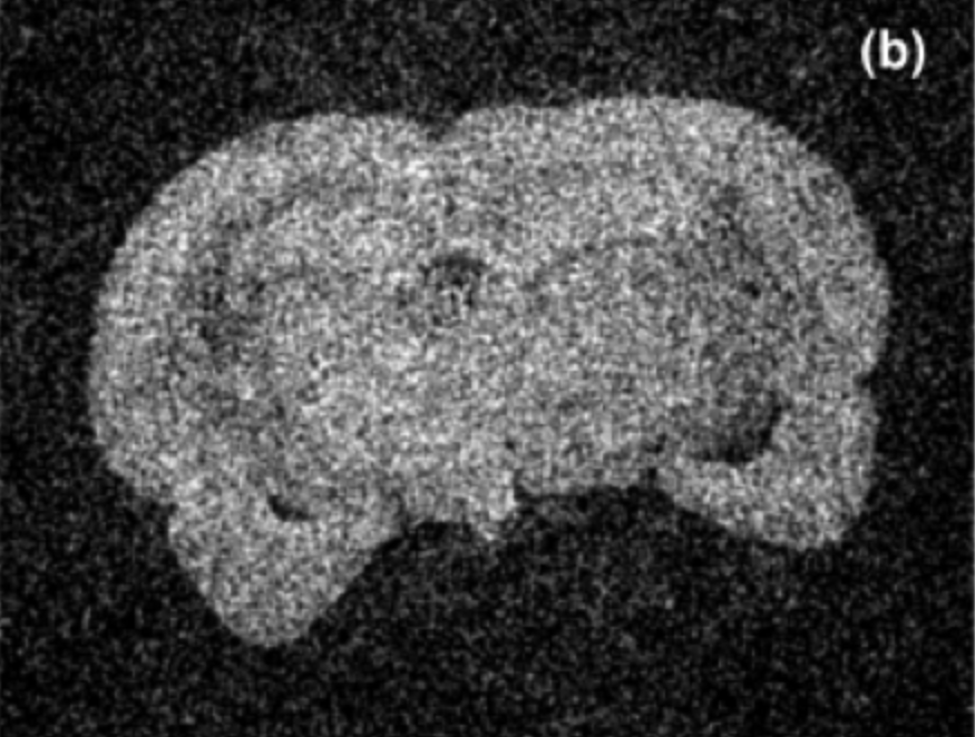
X-ray video of a breathing mouse acquired using an Excillum MetalJet D2+ X-ray source. The frame rate is 30 Hz and effective pixel size 19 µm. Video from M. Preissner et al. “Application of a novel in vivo imaging approach to measure pulmonary vascular responses in mice”, Physiol. Rep.(2018).
High-resolution zebrafish muscle imaging with X-ray PBI tomography
By using a MetalJet tube, PBI tomography of an unstained whole zebrafish was performed. This shows the capability of capturing the low contrast sub-5 µm details with suffciently high contrast. The methodology paves the path for the non-invasive whole-body imaging at sub-cellular resolution for soft tissue research and small animal models, thus facilitate the deep understanding of muscular disease and assessing interventions.
The PBI tomographic axial (a) and sagittal (b) slices and the zoom-in ROI (c) as the black box in (b).
- Exposure time = 115 s/projection
- Voxel size = 0.733 µm
- Tube voltage = 40 kV, tube power = 24 W
- FDI- VHR CCD camera with Gadox scintillator
W. Vågberg, et al., “X-ray phase-contrast tomography for high-spatial-resolution zebrafish muscle imaging“, Sci. Rep. 5. 16625 (2015).
High resolution X-ray PBI lung tomography on whole mice at laboratory scale
The scientists at University of Göttingen demonstrated the high resolution PBI tomography on whole mice based on using a MetalJet source. With the optimized spectrum they demonstrated the applicability of PBI in local tomography for small animal imaging of lungs, in the presence of highly absorbing surrounding tissue at µm- level resolution. The result indicated the application of this imaging method at cellular level for even larger biological samples.
3-D rendering of the PBI tomogram of the soft tissue in the thorax.
- Exposure time = 10 s/ radiography
- voxel size = 5 µm
- Tube voltage = 70 kV, tube power= 100 W
- Timepix photon counting detector
M. Krenkel, et al., “Propagation-based phase-contrast tomography for high-resolution lung imaging with laboratory source“, AIP Adv. (2016).
Cellular and sub-cellular resolution X-ray PBI tomography on mouse brain cytoarchitecture
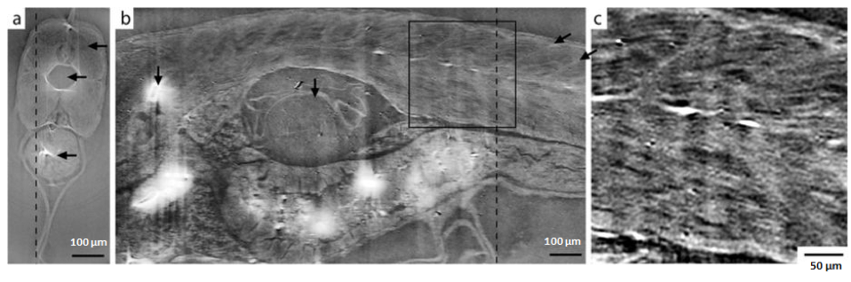
An another example from the research group in Göttingen more recently, the mouse brain cytoarchitecture was investigated at sub-celluar level resolution by PBI tomography. Together with suitable phase-retrieval algorithm and novel tissue prepartation, this study highlights that this is a non-destructive imaging method in the studies of brain architecture in mammals.

PBI tomography slice along transverse (a) and another along longitundnal (b) directions; automatic volume rendering of the reconstructed volume (c) and the zoomed-in cellular segmentation (d). The molecular layer (ML), granular layer (GL), white matter (WM) and Purkinje cell layer (PCL) of the cerebellar vermis at cellular resolution.
- Exposure time = 50 s/ radiography
- Voxel size = 47 µm
- Tube voltage = 40 kV, tube power= 50 W
- Lens-coupled scintillator-based camera XSight
M. Töpperwien, et al., “Three-dimensional mouse brain cytoarchitecture revealed by laboratory-based x-ray phasecontrast tomography“, Sci. Rep. (2017).
Grating-Based Phase-Contrast Imaging (GBI)
The core of GBI is the Talbot self-imaging effect: when spatially coherent X-rays pass through a grating (G1) with periodic line pattern, the line pattern will repeat itself at certain distances. By inserting a second grating (G2) that matches the self-image from G1, small differences introduced by a sample present in the beam can be detected. The second grating is shifted in several steps between where it is in phase and out of phase with the self-image from G1. This allows three properties of the object to be measured separately; X-ray attenuation, phase shift and dark field originating from ultra-small angle scattering. Separate attenuation and phase signals allow better discrimination of materials and the dark-field signal visualizes the presence of small structures below the imaging resolution.
By retrieving the changes in the self-image measured by stepping the G2 grating, the three image modalities can be measured simultaneously. GBI can often be implemented in a laboratory setup, but often requires a grating directly in front of the source, referred to as G0. The reason for this is that most sources don’t have the spatial coherence to enable image contrast in GBI, but each slit in the G0 grating works as a spatially coherent source but mutually incoherent to the other slits. Naturally, the G0 grating causes a loss in flux of more than 50%. A MetalJet X-ray source reaches the sufficient spatial coherence to enable GBI without the G0 grating, thus avoiding the loss of flux.
The most common way to acquire images in GBI is the phase stepping procedure that requires several images every projection. This has the downside that it associates GBI tomography to very long exposure times. In this context, it is of deepest concern to keep the flux high!
Application examples for Grating-Based Phase-Contrast Imaging (GBI)
X-ray GBI microscope with MetalJet source
The joint research by scientists from KTH and ETH/PSI proved the advantage of using a MetalJet source in GBI, which significantly improved flux and visibility compared to a conventional microfocus tube. Moreover, a preliminary exploration of its biomedical application has been shown by tomography on a rat.
A comparison between phase-contrast (a) and attenuation-contrast (b) slices from GBI tomography based on a MetalJet source. The sample was a rat brain scanned in a liquid paraffin bath.
- Exposure time = 1 min/ stepping image
- Voxel size = 20 µm
- Tube voltage = 50 kV, tube power= 40 W
- G1 is designed for π-phase shift at 25 keV, period= 4.12 µm
- CCD camera with Gadox scintillator
Reproduced from T. Thüring, et al., “X-ray grating interferometry with a liquid-metal-jet source“, Appl. Phys. Lett. (2013) with the permission of AIP Publishing.
X-ray grating interferometry at 9.25 keV with MetalJet tube
The research groups at Fraunhofer IIS (Würzburg, Germany) took the advantage of high brightness Gallium’s Kα line of MetalJet source and applied it to GBI, as well as the possibility of single-grating setup. The high visibility confirmed the high spatial coherence MetalJet source. Together with the high-resolution detector, a high spatial resolution of 2.7 µm has been achieved. Its application in materials science was further exploited.
By using the Gallium Kα line at 9.25 keV and high spatial coherence from a MetalJet source, researchers at the Fraunhofer IIS (Würzburg, Germany) show that grating interferometry is possible without the G0 grating. Furthermore, by employing a detector with very high resolution, the self-image from the G1 grating can be resolved, which eliminates the need for a G2 grating and phase stepping. This results in that all three image modalities can be captured in a simpler experimental setting. Here, the source was operated at 70 kV and 50 W, G1 grating a period of 2.4 µm and the detector had a pixel size of 0.67 µm.
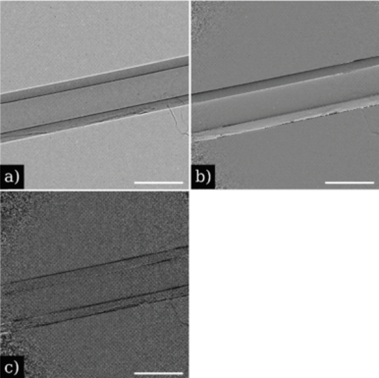
Single grating imaging with MetalJet source and a high-resolution detector. A nylon fiber is imaged and a single image captures(a) attenuation(b) phase and(c) dark field. Scalebar is 260 µm.
Reproduced from A. Balles, et al., “X-ray grating interferometry for 9.25 keV design energy at a liquid-metal-jet source“, AIP Conf. Proc. (2016) with the permission of AIP Publishing.
Speckle-Based Phase-Contrast Imaging (SBI)
SBI exploits the near-field speckle pattern generated by illuminating a random wave front modulator, also called diffuser, with spatially coherent X-rays. The multi-contrast images can be obtained by retrieving local image distortions of the acquired patterns due to the presence of the sample. The local distortions carry the information for absorption, phase shift and scattering of the sample. The resolution of SBI can be improved by doing so called speckle scanning, which consists of a 1D or 2D raster scan, but consequently needs longer acquisition time.
In similarity to GBI, a MetalJet source contributes to SBI by providing high spatial coherence to resolve the µm-sized speckles and high brightness to reduce exposure time.
Application examples for Speckle-Based Phase-Contrast Imaging (SBI)
Laboratory-scale SBI imaging based on MetalJet and scanning technique
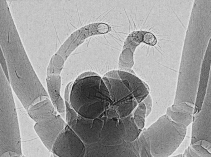
Thanks to the high-brightness MetalJet source, the scanning technique was demonstrated the first time for a compact SBI setup by scanning a phantom and a dried spider. A 7 µm fine hair is clearly visible in the retrieved images with a resolution higher than what can be achieved by the speckle-tracking method.
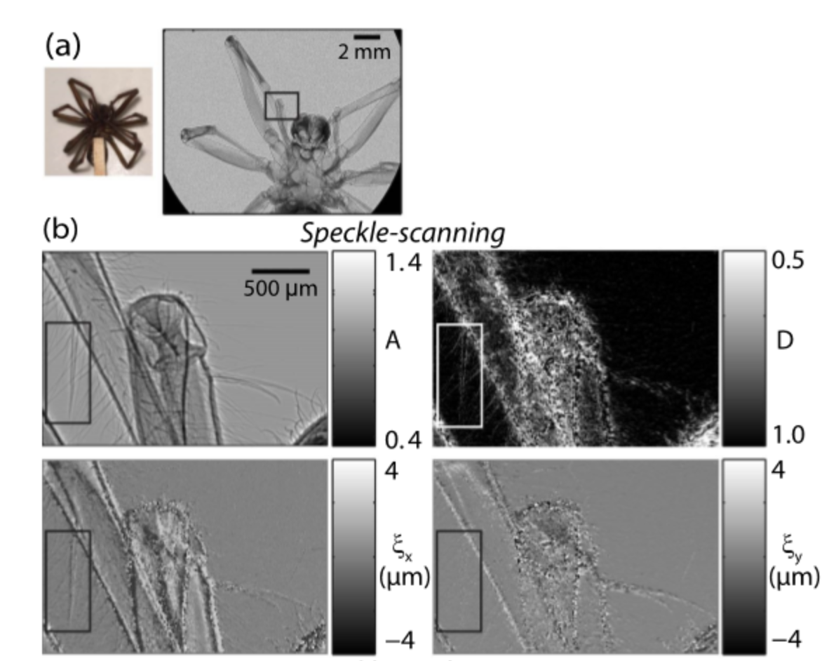
The radiographs of a dried spider in attenuation-contrast, dark-field contrast and phase-shift along transverse and longitudinal directions (b). (a) is the photo of the sample and one raw speckle image.
- Exposure time = 1 min/ stepping image
- Effective pixel size = 4.5 µm
- Tube voltage = 50 kV, tube power= 30 W
- 32 x 32 2-D raster scan with 1 µm step size
- CCD camera FLI PL 9000
T. H. Zhou, et al., “Speckle-based x-ray phase-contrast imaging with a laboratory source and the scanning technique“, Opt. Lett. (2015).
Laboratory-scale X-ray SBI microtomography for quantitative material characterisation
The collaboration between KTH and Diamond Light Source showed the possibility of doing quantitative material characterization using the attenuation-contrast and phase-contrast 3-D images acquired by SBI tomography based on a MetalJet source. The clear distinction among different material phases in the polymer colloidal suspension was demonstrated.
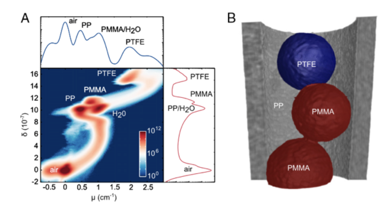
Quantitative analysis on the attenuation-contrast and phase-contrast tomograms on multi-material phantoms, which were acquired by the SBI tomography based on MetalJet source. The 2D histogram of voxel value on those two tomogram (a) and the 3D rendering of the tomogram (b).
- Exposure time = 2 min/ radiography
- Voxel size = 7 µm
- Tube voltage = 50 kV, tube power= 30 W
- FDI- VHR CCD camera with Gadox scintillator
I. Zanette, et al., “X-ray microtomography using correlation of near-field speckles for material characterization“, PNAS (2015).
Unless otherwise stated, pictures and content is published under license for CC-BY (https://creativecommons.org/licenses/by/4.0/).
Related user stories
Experiences with the Excillum X-ray sources
Learn about some of our customers' experiences with our state-of-the-art X-ray sources.