How 3D virtual histology based on µCT helps to understand Covid-19
In this talk, Prof. Salditt will present the current status of the project, the pathological relevance, and the technical challenges concerning data acquisition, reconstruction and analysis. He will finish with an outlook on how 3D virtual histology and patho-histology can be further developed and be applied to biomedical research, with an emphasis on compact laboratory instrumentation.
The webinar is 55 minutes.
Speaker
Prof. Dr. Tim Salditt
Professor of Experimental Physics, head of research group Structure of Biomolecular Assemblies and X-ray Physics, Institute of X-ray physics, University of Göttingen
Severe progression of Covid-19 is frequently accompanied by lethal respiratory failure. The underlying lung injury can already be detected by radiographic chest imaging and clinical computed tomography (CT). However, in order to study disease mechanisms at the cellular level, a much higher resolution is required. For this purpose, the tissue obtained by surgical intervention or from a postmortem autopsy is cut into thin sections, stained and observed in an optical microscope. In conventional histology, images are obtained only of two-dimensional sections but not of the entire three-dimensional (3D) volume. In order to accurately track the tree of blood vessels, to visualize alveolar morphology and to perform precise measurements of alveolar wall thickness, which is important for gas exchange in the lung, the cyto-architecture of the lung has to be visualized in 3D and at high resolution, after locating regions of interest such as inflammation sites. For this purpose, phase-contrast (PC) X-ray computerized tomography (CT) have been used such that three-dimensional imaging tasks can be performed non-destructively on lung autopsies or biopsies. The team at the University of Göttingen has implemented this both with synchrotron radiation, and at their in-house MetalJet source to investigate tissue samples from Covid-19 patients, as well as healthy control samples [1].
Using multi-scale phase contrast X-ray tomography, the team can augment the pathological assessment and understanding of Covid-19 pathophysiology based on detailed 3D visualizations of diffuse alveolar damage (DAD) with its prominent hyaline membrane formation, mapping out the distribution of immune cells infiltrating the tissue, and by providing histograms of characteristic distances from tissue interior to the closest air compartment. Most recently, the team have extended this to investigations of heart tissue, which can also show severe damage of vasculature.
[1] Marina Eckermann, Jasper Frohn, Marius Reichardt, Markus Osterhoff, Michael Sprung, Fabian Westermeier, Alexandar Tzankov, Christopher Werlein, Mark Kühnel, Danny Jonigk, Tim Saldittt, 3D virtual pathohistology of lung tissue from Covid-19 patients based on phase contrast X-ray tomography. eLife 2020;9:e60408 doi: 10.7554/eLife.60408
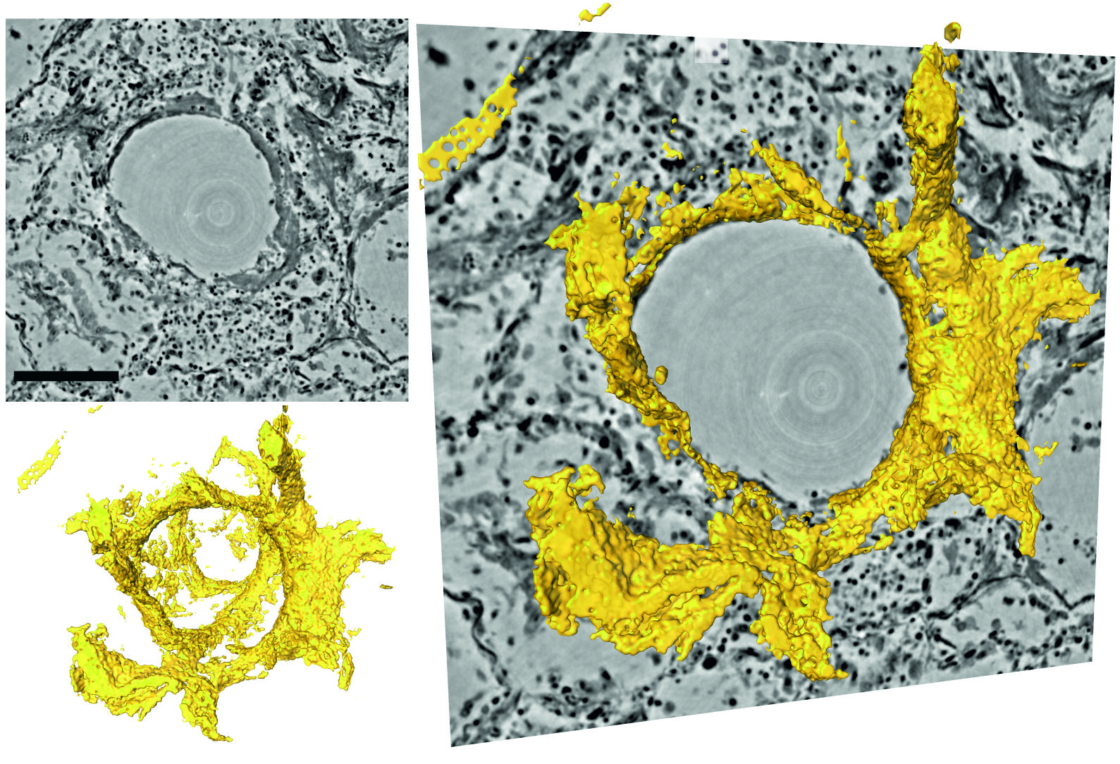
Covid-19 can lead to severe lung damage.
The alveolar walls (tiny air sacs) can be clogged with protein and cellular debris resulting from the inflammatory response. This so-called hyaline membrane, which is here visualized in 3D, can reduce or impede the gas exchange between the air compartment and the tiny neighbouring capillaries. Here, virtual slices through the reconstructed volume around an alveolus are shown, with superimposed rendering of the hyaline membrane attached to alveolar walls. Scale bar 300µm. (Image, adapted from [1])
Please register below.