A new dimension in cellular scanning
Professor Tim Salditt is not only a pioneer in phase-contrast biomedical imaging. He is also one of the first to implement the Excillum MetalJet in laboratory research, more than a decade ago. Professor Salditt and his team see enormous potential to create pre-clinical laboratory instruments for color CT scanning of tissue biopsies, based on their latest X-ray source: the Excillum NanoTube N2.
Interviewee
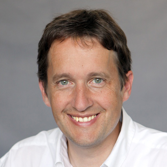
Prof. Dr. Tim Salditt, Professor of Experimental Physics, head of research group Structure of Biomolecular Assemblies and X-ray Physics
Institute
Institute of X-ray physics, University of Göttingen
Method
Phase-contrast imaging
Application
Cellular-scale tissue scanning
X-ray sources
MetalJet R5 (2009), MetalJet D2 (2013), NanoTube N1 (2017) and NanoTube N2 (2020)
With both sources it’s clear that we can really make the full transition from absorption-based imaging to phase-contrast imaging for very challenging samples. And this is what we want to continue, to push the limits of the technology and the application.
Prof. Dr. Tim Salditt
More information from every photon
Professor Salditt’s research group at the University of Göttingen is devoted to describing biological matter – from biomolecular systems to cells and tissues – in quantitative physical terms. To better understand the structures underlying biological functions, often at the nanoscale, the team is continuously developing the capabilities of nano-focused X-ray beams and phase-contrast X-ray tomography. To paraphrase the group’s goals in simpler terms, each experiment aims to get the maximum information from every photon, a pursuit that has perfectly aligned with Excillum’s own ambitions.
The Excillum sources remain a key to this endeavor. In particular, what we like is that they have a very high-quality electron source with a lot of controls. The hardware/software interface is also well done.
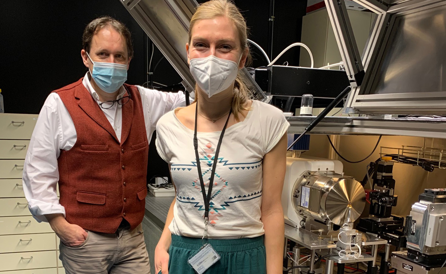
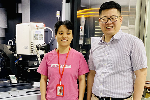
Prof. Dr. Tim Salditt, Professor of Experimental Physics (left) and PhD candidate Marina Eckermann (right) in front of the Excillum NanoTube N2 set-up in January 2021.
Professor Salditt’s original interest in the Excillum MetalJet, starting in 2009, stemmed from synchrotron-related research in phase-contrast imaging. “In our research,” he explains, “we have to scan various tissues at high resolution, and we are also deeply engaged in the development in phase-contrast techniques. So, the MetalJet was really an opportunity to scan samples before going to the synchrotron. There’s so much work in selecting, preparing and embedding samples, so we saw great potential with compact sources. The Excillum sources remain a key to this endeavor. In particular, what we like is that they have a very high-quality electron source with a lot of controls. The hardware/software interface is also well done.”
So, what new capabilities did the MetalJet offer? “The MetalJet gave us a phase-contrast capability at, let’s say, a few microns resolution,” says Salditt. “So it’s maybe not for the highest resolution, but it gives us a very good contrast. One of the big open questions in our research is how to best configure the source, the detector, the geometry and the reconstruction algorithms. This is a lot of what we do: we see a lot of gain in quality in matching these quantities. There are also good arguments to go to, let’s say, sub-micron focal sizes, adapt the detector geometry and then use that for tissues. It’s not only out of a desire to get a higher resolution from the small spot size, but also to improve the partial coherence and the visibility of details in our samples — hence the contrast.”
NanoTube and the future of 3D histology
These early experiences in enhancing image contrast with the MetalJet led to a continued collaboration using an early version of the Excillum NanoTube N1. This source made it possible to achieve high-quality images of neuronal tissues, the results of which were published in the Journal of Medical Imaging and presented at conferences by first author Marina Eckermann, PhD. Student in the Salditt lab. “We could see that we could get even faster scans as well as a better picture of what we could achieve with both modalities,” says Salditt. “With both sources it’s clear that we can really make the full transition from absorption-based imaging to phase-contrast imaging for very challenging samples. And this is what we want to continue, to push the limits of the technology and the application.”
In the fall of 2020, Professor Salditt’s team decided to upgrade their lab with the Excillum NanoTube N2, which combines the highest possible resolution with a higher X-ray brightness, higher level of automation and higher spot stability, as well as improved electron beam control. The following months will be dedicated to pairing the new source with the lab’s most recent detector prototype. “Now we feel we can fully utilize this technology with the NanoTube N2 upgrade because many things are so much easier in terms of the control and handling it well,” says Salditt. “Our next big story is how to bring in our latest detector prototype with the special advantages of the NanoTube N2. And the keyword here is color CT. Because the color CT scanning we all know from the hospital is absorption-based, and we think that color CT and phase contrast would also be a very good match — not for organ imaging or breast cancer tumors, but more for neuroscience and biomedical studies on a cellular level in tissue.”
Our next big story is how to bring in our latest detector prototype with the special advantages of the NanoTube N2. And the keyword here is color CT. Because the color CT scanning we all know from the hospital is absorption-based, and we think that color CT and phase contrast would also be a very good match — not for organ imaging or breast cancer tumors, but more for neuroscience and biomedical studies on a cellular level in tissue.
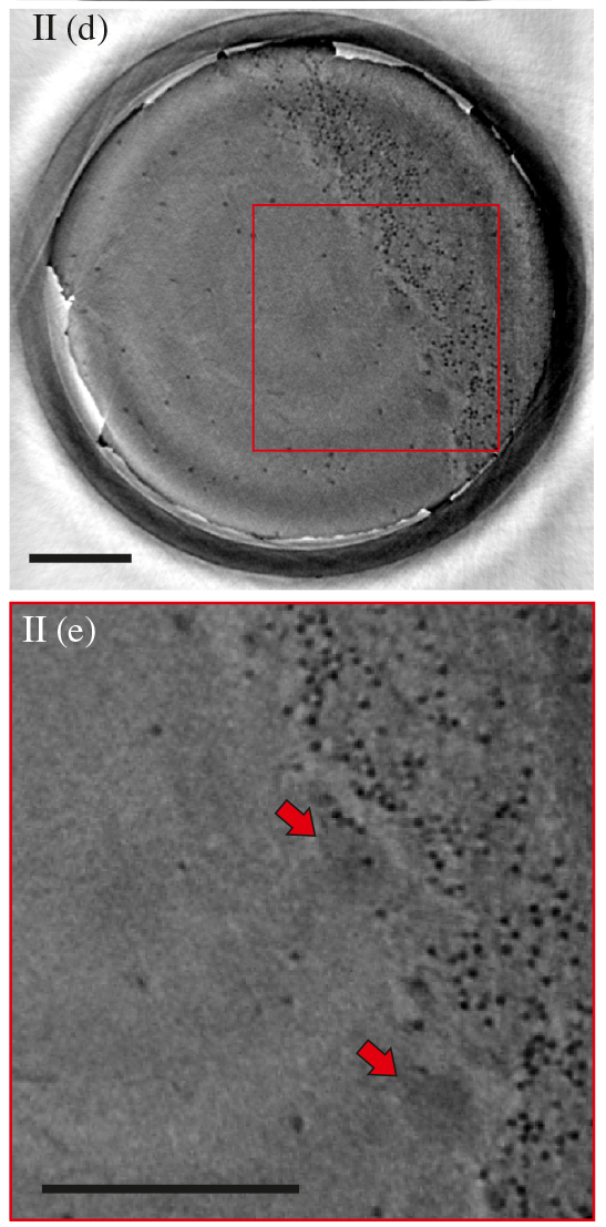
Slices from 3D virtual histology of neuronal tissue acquired using the NanoTube with a 0.5 µm spot size. Figure (d) shows an overview of the tissue while (e) is a zoom-in of the area marked in red in (d). Red arrows in (e) indicates Purkinje cells (PC). Scale bars: 100 µm.
The new setup, based on the NanoTube N2, is something Professor Salditt sees as a natural progression of their current research into visualizing tissue damage in severe Covid-19 cases, which includes a recent study published in the research journal eLife. “We’ll probably use this instrument to have a closer look at the Covid-19 lung and heart again,” he explains. “This is research that we’ve started this year (2020), and in that case it was the MetalJet that helped us to prepare the samples for our work with synchrotron radiation. Now we’ll also do this with the NanoTube N2, in particular for samples where you take a lung tissue, cultivate it and deliberately infect it with SARS-CoV-2 and see what goes wrong in a dish. We want to see: do we get the same damage without an immune system? What part of the damage is only due to the reproduction of the virus in the tissue? Because our collaborators can cultivate the tissue and use it as a test bench over a few days. Then we’ll stop at different time frames and look at it, and this has to be at high resolutions. Ultimately, we’ll be able to see the virus as it’s entering, but this is something that we’ll want to reach with synchrotron radiation. But I think that, say, a 300 nm voxel scan with the NanoTube N2, with the right detector, could also inform us very well already on the cellular scale.”
Phase-contrast tomography and pathology
These capabilities, according to Professor Salditt, make phase-contrast tomography especially well-suited for pathology, where the preparations of biopsy sections for optical microscopy are currently labor-intensive and often result in low-resolution output. “They have to do this very quickly, always sectioning in thin slices, and only in two dimensions. But many tissue structures can only be assessed if you track them in 3D. With small blood vessels, for instance, you need to see the vascular tree, the bifurcations of vessels, and it’s much too laborious to do this in slicing. It’s literally thousands of slices, and the resolution is also not sufficient. You really have to do it by tomography, and this is why I think there’s a real potential for advances in biomedical applications.”
We’ll probably use this instrument to have a closer look at the Covid-19 lung and heart again.
In the near future, Professor Salditt and his team see a significant role to play for phase-contrast tomography systems in enabling advanced 3D scanning of human tissues within pathology units — something that could lead to remarkable new insights into the diagnosis and treatment of diseases. “We believe that in each pathology unit of a university medical center, let’s say ten years, there should be a phase-contrast tomographic tissue scanner. This would histo-pathology of autopsies, but not only that. Biopsies, i.e. tissue samples of living people could also be analyzed for diagnostic purposes. And here I would probably see an instrument based on the NanoTube, rather than the MetalJet. It really depends, since there could still be configurations where you need to have a very fast scan, not the highest resolution. But you want a result very quickly if, for instance, the patient is still in surgery and you have only half an hour for a diagnostic input. If there were any case where you really need the three-dimensional output and a rather fast result, it could be an advantage to use a MetalJet. But so far, we’re very surprised by how much we can compensate for the lower power (of the NanoTube versus the MetalJet source) by moving the sample very close. And if the source spot is too large you can’t do this because you lose resolution. But if you shrink the source size — and the flux density is one over distance squared — then you really win by downsizing. We can have our biopsy samples at the cross section of 500 microns. We can go very close, and then it’s very advantageous.”
From live tissue to digital quantification
Recently (Dec 2020), Salditt’s lab has hired a new Phd student whose main focus will be to bring together the new NanoTube with the new detector technology. Asked if he has any advice to other researchers in similar biomedical imaging fields, Salditt replies, “We’ve been trying a lot in imaging and the main message is: It really works. The throughput and the information is there, and it’s this mixture of contrast, resolution and three-dimensionality that is crucial. One thing that really can’t be stressed strongly enough is that you get fully digitalized data that lends itself to quantification. So for the next ten years the real challenge will be in data science. We see so much data coming in and so much progress in algorithms. With tomography you get a large volume directly scanned into the computer, and you can have this high throughput, one after the other.”
This very challenge, to create both larger 3D overviews and close-up reconstructions of complex cellular structures, remains central to the lab’s mission. With the help of new sensors, new data science and innovative new X-ray sources, Salditt and his lab will continue to develop new methods, contributing to a better understanding of the relation between microscopic tissue structures, the larger organizational structure of organs and the targeted interventions and drugs that can improve human health.
We’ve been trying a lot in imaging and the main message is: It really works. The throughput and the information is there, and it’s this mixture of contrast, resolution and three-dimensionality that is crucial.