Enhancing biomedical research through X-ray phase-contrast imaging
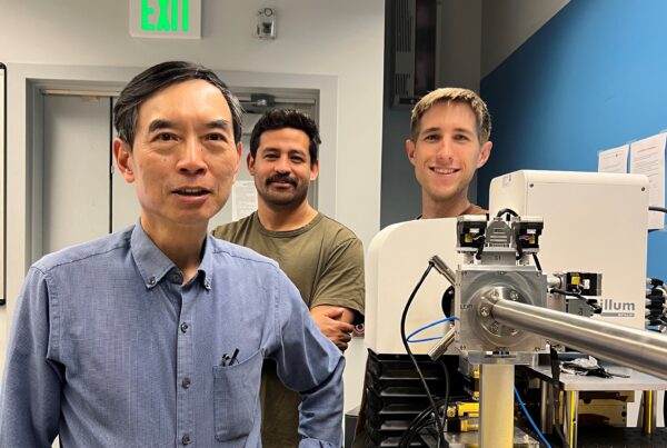
At Monash University in Melbourne, Australia, X-ray scientists are at the forefront of new techniques in phase-contrast imaging. In recent years, an in-house instrument based on the Excillum MetalJet has enabled a compact, simple to operate imaging set-up in comparison to synchrotron and compact light source methods.
Interviewees
Dr. Kaye Morgan and Dr. Isaac Pinar
Institute
Monash University, Melbourne, Australia
Method
X-ray phase-contrast imaging
Application
Biomedical research
X-ray sources
Excillum MetalJet D2+
Whereas a typical CT scan only lasts three to five minutes, some of these devices we end up testing for weeks. So in that case, you need a continuously running source that’s able to manage that imaging demand. No other sources really last that long. That’s a pretty clear benefit for us.
Dr. Isaac Pinar
For the biomedical researchers at Monash University, the requirements for advanced X-ray imaging are wide-ranging. “Our lab is primarily looking at cardiovascular-based devices,” says Dr. Isaac Pinar. “We use X-ray sources to look at everything surrounding that, whether it’s blood clotting in a cannula, for use in a clinic, an artificial valve or an oxygenator. In all of these areas, we’re trying to improve patient outcomes in terms of reducing blood clotting and adverse effects.”
One of the key challenges faced by the research team is that processes such as blood flow can be extremely difficult to simulate. The viscosity and flow of a particular fluid can be simulated, but their interactions with biological factors add multiple layers of complexity. Current methods for testing these medical devices, Isaac says, typically use laser techniques that require transparent materials. “That’s a huge change when you start considering how the biology in the blood interacts with the device’s surface. That’s why we’ve really shifted to looking at flows using X-rays of various sorts, to be able to see the device – and the flows passing through it – in its natural implantable material.”
From the synchrotron to the laboratory
Initially, the lab’s MetalJet source was tailored for use as a phase-contrast imaging system for lung motion tracking in small animals, led by Professor Andreas Fouras and Dr. Stephen Dubsky. Previous work had all been completed at a synchrotron X-ray source overseas, which limited the number and range of possible studies. Recent publications from Dr. Murrie and Dr. Werdinger have shown that the synchrotron techniques can also be performed with the MetalJet set-up.
According to Isaac, the high flux and cone beam produced by the MetalJet source enabled a system in which “you can image with short exposures because you’ve got the magnification to allow the use of larger pixels. This means you can use a thicker scintillator, which is much more efficient. You can collect enough photons for an image in a much shorter time than you would be able to with a low-divergence source that requires a detector with small pixels.”
Analyzing disease progression
The capabilities of this flexible set-up provide new opportunities for research studies that seek to better understand how the body works. Compared to the synchrotron experiments that Dr. Morgan and her team performed, the new instrument provides a lab-scale imaging system that is more suitable for researching diseases in live animals, where the effectiveness of new treatments can be studied in vivo and in real time using non-invasive x-ray imaging.
“It’s always there so you can do a lot of studies,” Dr. Morgan explains, “and you can image spontaneously when you get to some point of health with the animals. My colleagues and I were doing a study where we were looking at tumors growing in the lungs, and with the model we didn’t know if it was going to take two days or two weeks. You can’t plan a synchrotron experiment around that. But if you’ve got a source that’s capable of doing synchrotron experiments that you can always use, then you can do these kinds of things where you’re following the disease progression. That’s probably the key advantage: having your own beam line.”
In short, the new MetalJet set-up has made it possible to bridge a large capability gap and move synchrotron x-ray imaging advances towards human diagnostics, evidence of which can already seen at Monash with Professor Fouras’ company 4Dx, which is now commercializing its lung motion tracking technology and testing it in hospitals.
A broad range of sample sizes
The availability of an in-house beam line has also given the lab the freedom to tailor the system to a wide range of specimens and samples. “To give you an idea,” says Isaac, “we’ve started imaging some pretty cool stuff. We’ve gone from imaging blood flow through brains to arthritis in mice.” To handle all of these different applications, the research team has naturally refined and adapted the system over time. “The magnification and all sorts of things have needed to change,” he continues, “so it’s a lot more flexible at the moment.”
The particular devices being tested in the lab also need to be tested for relatively long durations. Whereas a typical CT scan only lasts three to five minutes, says Isaac, “some of these devices we end up testing for weeks. So in that case, you need a continuously running source that’s able to manage that imaging demand. No other sources really last that long. That’s a pretty clear benefit for us.”
My colleagues and I were doing a study where we were looking at tumors growing in the lungs, and with the model we didn’t know if it was going to take two days or two weeks. You can’t plan a synchrotron experiment around that. But if you’ve got a source that’s capable of doing synchrotron experiments that you can always use, then you can do these kinds of things where you’re following the disease progression. That’s probably the key advantage: having your own beam line.
In the near term, the Monash University research team hopes to continue to enhance the flexibility of their phase-contrast imaging system. Whether the samples are brains, knees or valves, says Isaac, “my aim is to have the system flexible enough to accommodate all the sample sizes and effective pixel sizes that we need to achieve.”