Author: Dr. David Bernard, X-ray Consultant for the Electronics Industry (dbc@bernard.abel.co.uk)
This 2-part article has been commissioned by Excillum to celebrate the 125th anniversary of the discovery of X-rays on 8 November 1895. The first part reviews the history of X-rays and their use from then to now. The second looks at the history and recent developments in X-ray sources and show how their capabilities and use continues to grow and impact on our lives.
Part 1 – Early years
X-rays are a penetrating form of high energy electromagnetic radiation that are located between the UV and gamma rays in the electromagnetic spectrum. They were first discovered on 8 November 1895 when Wilhelm Conrad Röntgen stumbled across their effects to create images on fluorescent screens, and subsequently on photographic plates, at a distance from an optically hidden Crookes, or similar, tube. This would mean that X-rays would have been produced by experimenters from as early as 1875, when Crookes tubes, and their like, were first created.
However, it was Röntgen that first saw their effect and which he described in his paper “On A New Kind of Rays” that was published on 28 December 1895. Röntgen called his discovery X-rays to indicate that it was, then, an unknown type of radiation. However, in many languages to this day they are still known as Röntgen rays.
This discovery was recognised in 1901 when Röntgen was awarded the first Nobel prize for Physics. Since the discovery of X-rays, their use for analysis and diagnostics have been some of the most widely researched areas in science and engineering. It is amazing to note that in the first quarter of the 20th century, almost half of the Nobel prizes awarded were connected to contributions in this area.
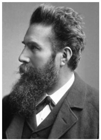
Wilhelm Conrad Röntgen 27/3/1845 – 10/2/1923 (image from www.nobelprize.org)
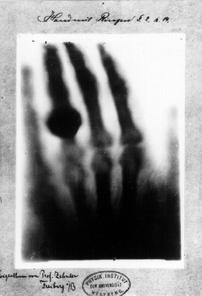
Print of first ‘medical’ X-ray image of Anna Röntgen’s hand that shows her wedding ring. (Ref: Kevles, Bettyann & Holtzmann (1996). ‘Naked to the Bone Medical Imaging in the Twentieth Century’. Camden, NJ: Rutgers University Press. pp. 19–22. ISBN 978-0-8135-2358-3)
Röntgen was quick to see the medical implications and opportunities of his discovery, as the famous first X-ray image of his wife Anna’s hand on 22 December 1895 shows. He realised that X-rays provided a non-invasive method to investigate inside the body. It is interesting to note that, for personal ethical reasons, Röntgen refused to take out patents on his discovery of X-rays, as he wanted society, as a whole, to benefit from their practical applications. As a result, this quickly led to an explosion in research papers and development into X-rays and X-ray equipment all over the world, in a similar way that may be more familiar to us today in what happened after the initial discovery of graphene.
Broad medical usage of the newly discovered X-rays was widespread within only a few years. X-ray investigations were widely used in the first world war, where even the existing, and future, Nobel prize winners, and mother and daughter, Marie and Irène Joliot-Curie operated mobile and fixed X-ray units for wounded soldiers in Belgium.
Quickly following on from the diagnostic medical applications of the newly discovered 2D X-ray images came the use of X-rays for therapy, the developments of which are seen and used today for the treatment of cancers through radiotherapy and brachytherapy. Outside of the medical field, X-ray sources were being used for other, new techniques enabling fantastic new discoveries. These techniques include X-ray diffraction and Compton back-scattering and fluorescence methods. So, for example, it is only 58 years from the date of the first X-ray image to when X-rays were used to confirm the structure of DNA through X-ray diffraction. X-ray fluorescence and diffraction continue to be vital for materials analysis today.
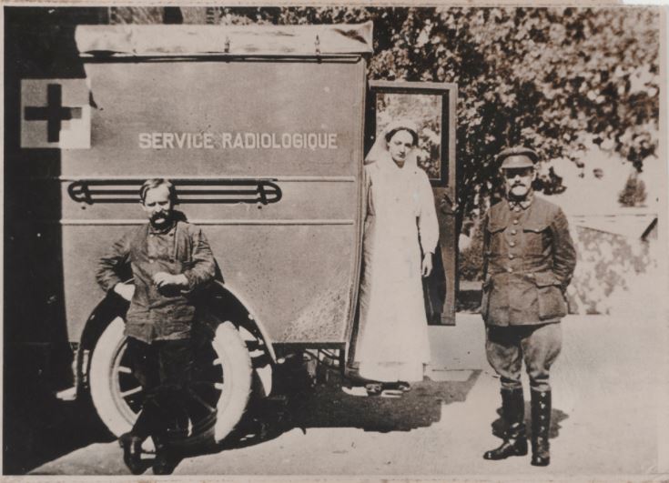
Irène Joliot-Curie climbing down from a “radiological car”, 1916 © Association Curie Joliot-Curie (or can reference through www.nobelprize.org, where it is shown).
X-ray microscopy
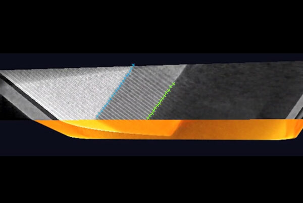
Outside of medical applications, another of the most far reaching developments for new uses of X-rays came in 1951 when Cosslett & Nixon invented the X-ray (shadow) microscope at the University of Cambridge (see image) (Ref. X-ray Microscopy, Cosslett V E and Nixon W, Cambridge University Press, Cambridge (UK), 1960). They showed that moving an object closer to the focal spot of an X-ray source, when there is a fixed detector position, creates an increasingly magnified (shadow) image of the object at the detector. The geometric magnification of the resultant image is calculated by the ratio of the, distance of the source focal spot to the detector, to the distance of the source focal spot to the object. Most industrial X-ray equipment follow this design concept so that good imaging is accompanied by an acceptable level of magnification for the investigation task. This is different from typical medical applications where the object (e.g. a limb) is placed on the detector (e.g. a film) and so provides an unmagnified image. X-ray microscopy has allowed practical and multi-industry usage for non-destructive investigation and quality control in such areas, amongst others, as:
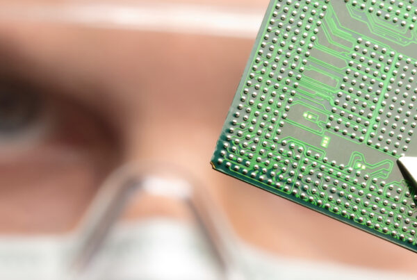
- The manufacture of ever-shrinking electronic components and circuit boards.
- Issues in food security, such as the identification of possible contamination from the raw materials or the production equipment.
- Thickness and defect monitoring over the whole range of non-destructive testing applications, such as for castings and pipe welds.
- Quality and safety control in tyre manufacture.
- Pharmaceutical quality control.
- Security applications.
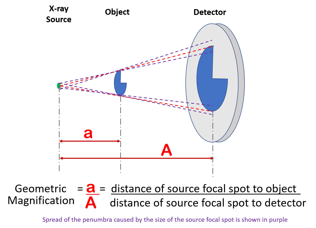
Schematic of X-ray (shadow) Microscope showing how to calculate the geometric magnification of an object image when seen at the detector.
Godfrey N. Hounsfield, 28/8/1919 – 12/8/2004 (Ref: image from www.nobelprize.org)
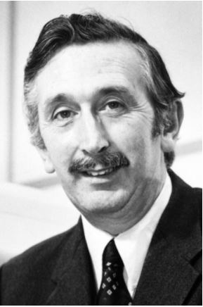
The 2D X-ray image, with or without magnification, has provided fantastic benefits since its discovery. The next major development was in the late 1960s when Godfrey Hounsfield added a third dimension for X-ray analysis with the discovery of Computer Assisted Tomography, now generally known as CT. Not only has this provided the ability to non-destructively slice through different layers of an object with, or without, magnification, whether it is medical or industrial in nature, it is arguable that it also spurred on other novel imaging techniques such as Magnetic Resonance Imaging, or MRI, that has revolutionised diagnostic medicine in recent years. Hounsfield shared the 1979 Nobel Prize in Physiology or Medicine with Allan Cormack for the discovery of CT. The invention of the MRI won the Nobel Prize for Physiology or Medicine in 2003.
By adding a third dimension to X-ray analysis, not only has CT enhanced the capabilities of medical diagnostics and helped locate and better target tumour treatment, but it has also helped existing and new industrial applications such as:
- Prospecting for new sources of oil by investigating the structure of rock samples.
- Analysing novel materials and composites.
- The investigation and quality control of additive manufacturing which are the industrial applications from 3D printing.
In the 125 years since Röntgen first glimpsed the effects of those ‘unknown’ rays not all uses of X-rays have flourished successfully. For example, the X-ray shoe fitting machine that was a common sight in shoe shops the USA between 1930 and 1950. However, the benefits and opportunities that X-rays have provided across a large range of industries and applications massively outweighs those small failures. The next part of this article will look in more detail at the history and developments in the X-ray sources themselves that have allowed all these X-ray applications to occur.
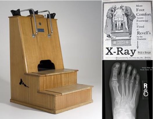
X-Ray Shoe-Fitting Machine common in the USA between 1930-1950 (Ref: www.americacomesalive.com)
Published: November 8, 2020